Trichophyton interdigitale: Difference between revisions
CSV import |
CSV import |
||
| Line 43: | Line 43: | ||
[[Category:Dermatophytes]] | [[Category:Dermatophytes]] | ||
[[Category:Fungal pathogens of humans]] | [[Category:Fungal pathogens of humans]] | ||
== Trichophyton_interdigitale == | |||
<gallery> | |||
File:Trichophyton_mentagrophytes_(257_18)_Cultured.jpg|Trichophyton interdigitale cultured | |||
File:Trichophyton_mentagrophytes_(257_19)_From_a_microculture_and_an_infected_hair.jpg|Trichophyton interdigitale from a microculture and an infected hair | |||
</gallery> | |||
Latest revision as of 02:17, 18 February 2025
Species of fungus
Trichophyton interdigitale is a species of fungus in the genus Trichophyton. It is a dermatophyte, which means it is a type of fungus that causes skin infections in humans and animals. T. interdigitale is closely related to Trichophyton mentagrophytes and is often associated with athlete's foot and other tinea infections.
Description[edit]
Trichophyton interdigitale is characterized by its ability to grow on keratinized tissues such as skin, hair, and nails. The fungus forms colonies that are typically white to cream-colored with a powdery texture. Microscopically, it produces septate hyphae and conidia. The conidia are usually spherical to pyriform and are borne singly or in clusters.
Pathogenicity[edit]
Trichophyton interdigitale is a common cause of dermatophytosis, particularly in the feet, where it causes tinea pedis (athlete's foot). It can also infect the nails, leading to onychomycosis, and occasionally other parts of the body, resulting in tinea corporis or tinea cruris. The infection is typically superficial, affecting the outer layers of the skin, but can cause significant discomfort and itching.
Diagnosis[edit]
Diagnosis of infections caused by Trichophyton interdigitale is usually made by clinical examination and confirmed by laboratory tests. Microscopy and culture of skin scrapings, nail clippings, or hair samples can reveal the presence of the fungus. Molecular techniques such as PCR can also be used for more precise identification.
Treatment[edit]
Treatment of Trichophyton interdigitale infections typically involves the use of topical or oral antifungal medications. Common topical treatments include terbinafine, clotrimazole, and miconazole. In more severe cases, oral antifungals such as itraconazole or fluconazole may be prescribed.
Prevention[edit]
Preventive measures include maintaining good personal hygiene, keeping the skin dry, and avoiding sharing personal items such as towels and footwear. In communal areas such as swimming pools and locker rooms, wearing protective footwear can help reduce the risk of infection.
Related pages[edit]
References[edit]
- Weitzman, I., & Summerbell, R. C. (1995). The dermatophytes. Clinical Microbiology Reviews, 8(2), 240-259.
- Havlickova, B., Czaika, V. A., & Friedrich, M. (2008). Epidemiological trends in skin mycoses worldwide. Mycoses, 51(Suppl 4), 2-15.
Trichophyton_interdigitale[edit]
-
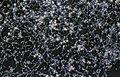 Trichophyton interdigitale cultured
 -
 Trichophyton interdigitale from a microculture and an infected hair
_Cultured.jpg)
_From_a_microculture_and_an_infected_hair.jpg)
