X-ray image intensifier
An X-ray image intensifier (XRII) is a medical imaging device that enhances the visibility of the internal structures of the body using X-rays. It is a key component in various diagnostic and therapeutic procedures, including fluoroscopy, radiography, and angiography. By converting X-rays into a visible light image, X-ray image intensifiers allow for real-time observation and analysis of bodily functions, structures, and pathologies.
History[edit]
The development of the X-ray image intensifier began in the early 20th century, following the discovery of X-rays by Wilhelm Conrad Röntgen in 1895. The first practical XRII was introduced in the 1950s, marking a significant advancement in medical imaging technology. Over the decades, improvements in image quality, reduction in radiation exposure, and the integration of digital technologies have greatly enhanced the functionality and applications of XRII.
Function[edit]
The primary function of an X-ray image intensifier is to increase the brightness of the image produced by X-rays passing through the body. This process involves several key components:
- Input Phosphor Screen: Converts incoming X-ray photons into visible light photons.
- Photocathode: Converts the light photons into electrons.
- Electrostatic Lenses: Focus and accelerate the electrons.
- Output Phosphor Screen: Converts the electrons back into a bright, visible light image.
This intensified image can then be viewed directly by medical personnel or captured by a camera for digital processing and display.
Applications[edit]
X-ray image intensifiers are utilized in a variety of medical procedures, including:
- Fluoroscopy: Real-time imaging of the movement of internal tissues, organs, or devices.
- Angiography: Imaging of blood vessels to detect abnormalities or blockages.
- Cardiac Catheterization: Diagnosis and treatment of cardiovascular conditions.
- Orthopedic Surgery: Real-time imaging to guide surgical procedures.
Advantages and Limitations[edit]
Advantages:
- Enhanced Image Quality: Provides clearer, more detailed images for accurate diagnosis and treatment.
- Reduced Radiation Exposure: Increases the efficiency of X-ray photon conversion, requiring less radiation to produce high-quality images.
- Real-time Imaging: Facilitates immediate observation and decision-making during medical procedures.
Limitations:
- Cost: High acquisition and maintenance costs.
- Size and Mobility: Larger and less mobile than some newer digital imaging systems.
- Radiation Exposure: Despite reductions, still poses a risk of radiation exposure to patients and healthcare providers.
Future Developments[edit]
Advancements in digital imaging technology are leading to the gradual replacement of traditional X-ray image intensifiers with flat-panel detectors (FPDs). FPDs offer several benefits over XRIIs, including improved image quality, lower radiation doses, and more compact designs. However, XRIIs continue to be used widely due to their proven effectiveness and the high cost of transitioning to fully digital systems.
See Also[edit]
This medical article is a stub. You can help WikiMD by expanding the page. |
-
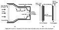 X-ray image intensifier schematic
 -
 Mobile X-ray image intensifier


Medical Disclaimer: WikiMD is for informational purposes only and is not a substitute for professional medical advice. Content may be inaccurate or outdated and should not be used for diagnosis or treatment. Always consult your healthcare provider for medical decisions. Verify information with trusted sources such as CDC.gov and NIH.gov. By using this site, you agree that WikiMD is not liable for any outcomes related to its content. See full disclaimer.
Credits:Most images are courtesy of Wikimedia commons, and templates, categories Wikipedia, licensed under CC BY SA or similar.
Translate page: - East Asian
中文,
日本,
한국어,
South Asian
हिन्दी,
தமிழ்,
తెలుగు,
Urdu,
ಕನ್ನಡ,
Southeast Asian
Indonesian,
Vietnamese,
Thai,
မြန်မာဘာသာ,
বাংলা
European
español,
Deutsch,
français,
Greek,
português do Brasil,
polski,
română,
русский,
Nederlands,
norsk,
svenska,
suomi,
Italian
Middle Eastern & African
عربى,
Turkish,
Persian,
Hebrew,
Afrikaans,
isiZulu,
Kiswahili,
Other
Bulgarian,
Hungarian,
Czech,
Swedish,
മലയാളം,
मराठी,
ਪੰਜਾਬੀ,
ગુજરાતી,
Portuguese,
Ukrainian
