Pyelogram
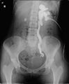
Pyelogram is a type of medical imaging procedure that allows physicians to visualize the anatomy and functionality of a patient's kidneys, ureters, and bladder. The procedure involves the injection of a contrast medium into the patient's bloodstream, which then travels to the kidneys and urinary tract, highlighting these areas on an X-ray image.
Procedure[edit]
The pyelogram procedure begins with the patient lying on an X-ray table. A radiologist or a trained technician will then inject the contrast medium into a vein, usually in the patient's arm. The contrast medium travels through the bloodstream and eventually reaches the kidneys, where it is filtered out and passes into the ureters and bladder. X-ray images are taken at various stages to visualize the kidneys, ureters, and bladder.
Types of Pyelogram[edit]
There are two main types of pyelogram: intravenous pyelogram (IVP) and retrograde pyelogram.
- Intravenous Pyelogram (IVP): This is the most common type of pyelogram. The contrast medium is injected into a vein and images are taken as the contrast medium is filtered by the kidneys and passes through the urinary tract.
- Retrograde Pyelogram: This procedure is used when an IVP is not possible or does not provide sufficient information. In a retrograde pyelogram, the contrast medium is injected directly into the ureters through a cystoscope, a thin tube inserted through the urethra.
Uses[edit]
Pyelograms are used to diagnose and monitor a variety of conditions affecting the kidneys and urinary tract. These include:
Risks[edit]
As with any medical procedure, there are potential risks associated with a pyelogram. These may include:
- Allergic reaction to the contrast medium
- Kidney damage in patients with poor kidney function
- Infection
- Bleeding
Patients should discuss these risks with their doctor before undergoing a pyelogram.
See Also[edit]

This WikiMD article can only be edited by registered and verified editors. You can log in or register.
Pyelogram[edit]
-
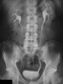 Pyelogram
 -
 Antegrade pyelogram of grade III hydronephrosis with obstruction at the ureterovesical junction


Medical Disclaimer: WikiMD is for informational purposes only and is not a substitute for professional medical advice. Content may be inaccurate or outdated and should not be used for diagnosis or treatment. Always consult your healthcare provider for medical decisions. Verify information with trusted sources such as CDC.gov and NIH.gov. By using this site, you agree that WikiMD is not liable for any outcomes related to its content. See full disclaimer.
Credits:Most images are courtesy of Wikimedia commons, and templates, categories Wikipedia, licensed under CC BY SA or similar.
Translate page: - East Asian
中文,
日本,
한국어,
South Asian
हिन्दी,
தமிழ்,
తెలుగు,
Urdu,
ಕನ್ನಡ,
Southeast Asian
Indonesian,
Vietnamese,
Thai,
မြန်မာဘာသာ,
বাংলা
European
español,
Deutsch,
français,
Greek,
português do Brasil,
polski,
română,
русский,
Nederlands,
norsk,
svenska,
suomi,
Italian
Middle Eastern & African
عربى,
Turkish,
Persian,
Hebrew,
Afrikaans,
isiZulu,
Kiswahili,
Other
Bulgarian,
Hungarian,
Czech,
Swedish,
മലയാളം,
मराठी,
ਪੰਜਾਬੀ,
ગુજરાતી,
Portuguese,
Ukrainian
