Purkinje images
Purkinje images are optical reflections that originate from the structures within the eye. They are named after the Czech anatomist Jan Evangelista Purkyně who first described them in 1823. These images are a series of bright spots that can be observed when light is reflected from the eye's surfaces. They provide insight into the optical properties of the eye and are used in various fields such as ophthalmology, optometry, and vision science.
Formation and Characteristics[edit]
Purkinje images are formed by the reflection of light from the different refractive interfaces within the eye. There are typically four recognized Purkinje images:
- Purkinje Image I: This image is formed by the reflection from the anterior surface of the cornea. It is the brightest and most easily observed of the four images.
- Purkinje Image II: This image results from the reflection off the posterior surface of the cornea. It is less bright than the first image and is inverted.
- Purkinje Image III: This image is created by the reflection from the anterior surface of the lens of the eye. It is dimmer than the first two images and is also inverted.
- Purkinje Image IV: The fourth image is formed by the reflection from the posterior surface of the lens. It is the dimmest and hardest to observe due to its location and the light absorption by the lens.
The relative positions and brightness of these images can change based on the angle of incoming light and the curvature of the eye's surfaces. The study of Purkinje images can reveal information about the eye's refractive health and has applications in diagnosing and understanding various eye conditions.
Applications[edit]
Purkinje images have several applications in medical and vision science:
- Eye Examination: They can be used to assess the shape and alignment of the eye's surfaces, aiding in the diagnosis of conditions like astigmatism or keratoconus.
- Measurement of Eye Movement: By tracking the movement of Purkinje images, researchers can study the dynamics of eye movements and understand the mechanisms behind them.
- Refractive Surgery: The analysis of Purkinje images can assist in planning and evaluating the outcomes of refractive surgeries such as LASIK.
Research[edit]
Research into Purkinje images continues to provide insights into the eye's optical properties and its health. Advances in imaging technology have made it easier to observe and analyze these reflections, leading to improved diagnostic techniques and a better understanding of the visual system.
See Also[edit]
References[edit]
This medical article is a stub. You can help WikiMD by expanding the page. |
-
 Diagram of four Purkinje images
 -
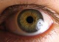 Pupil
Pupil


Medical Disclaimer: WikiMD is for informational purposes only and is not a substitute for professional medical advice. Content may be inaccurate or outdated and should not be used for diagnosis or treatment. Always consult your healthcare provider for medical decisions. Verify information with trusted sources such as CDC.gov and NIH.gov. By using this site, you agree that WikiMD is not liable for any outcomes related to its content. See full disclaimer.
Credits:Most images are courtesy of Wikimedia commons, and templates, categories Wikipedia, licensed under CC BY SA or similar.
Translate page: - East Asian
中文,
日本,
한국어,
South Asian
हिन्दी,
தமிழ்,
తెలుగు,
Urdu,
ಕನ್ನಡ,
Southeast Asian
Indonesian,
Vietnamese,
Thai,
မြန်မာဘာသာ,
বাংলা
European
español,
Deutsch,
français,
Greek,
português do Brasil,
polski,
română,
русский,
Nederlands,
norsk,
svenska,
suomi,
Italian
Middle Eastern & African
عربى,
Turkish,
Persian,
Hebrew,
Afrikaans,
isiZulu,
Kiswahili,
Other
Bulgarian,
Hungarian,
Czech,
Swedish,
മലയാളം,
मराठी,
ਪੰਜਾਬੀ,
ગુજરાતી,
Portuguese,
Ukrainian
