Posterior vitreous detachment

Editor-In-Chief: Prab R Tumpati, MD
Obesity, Sleep & Internal medicine
Founder, WikiMD Wellnesspedia &
W8MD's medical weight loss NYC, sleep center NYC
Philadelphia medical weight loss and Philadelphia sleep clinics
| Posterior vitreous detachment | |
|---|---|

| |
| Synonyms | PVD |
| Pronounce | N/A |
| Specialty | N/A |
| Symptoms | Floaters, flashes of light, visual impairment |
| Complications | Retinal detachment, macular hole |
| Onset | Typically after age 50 |
| Duration | Permanent |
| Types | N/A |
| Causes | Aging, myopia, trauma |
| Risks | Age, myopia, eye surgery |
| Diagnosis | Ophthalmoscopy, ultrasound |
| Differential diagnosis | Retinal detachment, vitreous hemorrhage |
| Prevention | N/A |
| Treatment | Usually none, vitrectomy if complications occur |
| Medication | N/A |
| Prognosis | Generally good, but complications can occur |
| Frequency | Common in older adults |
| Deaths | N/A |

Posterior Vitreous Detachment (PVD) is an eye condition where the vitreous membrane separates from the retina. It primarily occurs as a natural part of aging.
Pathophysiology[edit]
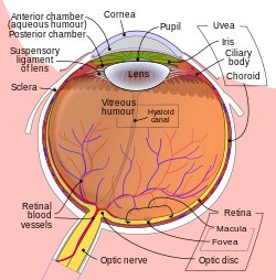
PVD involves the separation of the posterior hyaloid membrane from the retina posterior to the vitreous base, a 3-4 mm attachment to the ora serrata. The process is often due to changes in the vitreous humor consistency and volume with age.
Epidemiology[edit]
- Prevalence in Older Adults
Over 75% of individuals over the age of 65 experience PVD. The condition becomes increasingly common with advancing age.
- Occurrence in Middle-Aged Individuals
While less frequent in people in their 40s and 50s, PVD is not uncommon in this age group.
- Gender Differences
Some studies indicate a higher prevalence of PVD in women compared to men.
Clinical Features[edit]
Symptoms of PVD can include:
- Floaters
- Flashes of light
- A ring-shaped floater, indicative of a Weiss ring
Diagnosis[edit]
Diagnosis of PVD is primarily based on patient history and a comprehensive eye examination, including:
- Ophthalmoscopy
- Slit lamp examination
- OCT scans in some cases
Management and Prognosis[edit]
Most cases of PVD are benign and do not require treatment. However, patients should be monitored for complications like:
Patient Education[edit]
Patients with PVD should be educated about symptoms of retinal detachment and the importance of timely ophthalmologic evaluation if these symptoms occur.
References[edit]
- Johnson, M. W. (2010). Posterior vitreous detachment: Evolution and complications of its early stages. American Journal of Ophthalmology, 149(3), 371-382.
- Hikichi, T., Yoshida, A., & Akiba, J. (1995). Rate of posterior vitreous detachment in women with idiopathic macular hole. Archives of Ophthalmology, 113(6), 724-728.
See Also[edit]
Ad. Transform your life with W8MD's Budget GLP-1 injections from $75


W8MD offers a medical weight loss program to lose weight in Philadelphia. Our physician-supervised medical weight loss provides:
- Weight loss injections in NYC (generic and brand names):
- Zepbound / Mounjaro, Wegovy / Ozempic, Saxenda
- Most insurances accepted or discounted self-pay rates. We will obtain insurance prior authorizations if needed.
- Generic GLP1 weight loss injections from $75 for the starting dose.
- Also offer prescription weight loss medications including Phentermine, Qsymia, Diethylpropion, Contrave etc.
NYC weight loss doctor appointmentsNYC weight loss doctor appointments
Start your NYC weight loss journey today at our NYC medical weight loss and Philadelphia medical weight loss clinics.
- Call 718-946-5500 to lose weight in NYC or for medical weight loss in Philadelphia 215-676-2334.
- Tags:NYC medical weight loss, Philadelphia lose weight Zepbound NYC, Budget GLP1 weight loss injections, Wegovy Philadelphia, Wegovy NYC, Philadelphia medical weight loss, Brookly weight loss and Wegovy NYC
|
WikiMD's Wellness Encyclopedia |
| Let Food Be Thy Medicine Medicine Thy Food - Hippocrates |
Medical Disclaimer: WikiMD is not a substitute for professional medical advice. The information on WikiMD is provided as an information resource only, may be incorrect, outdated or misleading, and is not to be used or relied on for any diagnostic or treatment purposes. Please consult your health care provider before making any healthcare decisions or for guidance about a specific medical condition. WikiMD expressly disclaims responsibility, and shall have no liability, for any damages, loss, injury, or liability whatsoever suffered as a result of your reliance on the information contained in this site. By visiting this site you agree to the foregoing terms and conditions, which may from time to time be changed or supplemented by WikiMD. If you do not agree to the foregoing terms and conditions, you should not enter or use this site. See full disclaimer.
Credits:Most images are courtesy of Wikimedia commons, and templates, categories Wikipedia, licensed under CC BY SA or similar.
Translate this page: - East Asian
中文,
日本,
한국어,
South Asian
हिन्दी,
தமிழ்,
తెలుగు,
Urdu,
ಕನ್ನಡ,
Southeast Asian
Indonesian,
Vietnamese,
Thai,
မြန်မာဘာသာ,
বাংলা
European
español,
Deutsch,
français,
Greek,
português do Brasil,
polski,
română,
русский,
Nederlands,
norsk,
svenska,
suomi,
Italian
Middle Eastern & African
عربى,
Turkish,
Persian,
Hebrew,
Afrikaans,
isiZulu,
Kiswahili,
Other
Bulgarian,
Hungarian,
Czech,
Swedish,
മലയാളം,
मराठी,
ਪੰਜਾਬੀ,
ગુજરાતી,
Portuguese,
Ukrainian


