Middle cranial fossa
The middle cranial fossa, one of the three major depressions in the base of the skull, plays a critical role in both neuroanatomy and neurosurgery. It is deeper than the anterior cranial fossa and sits anterior to the posterior cranial fossa. The middle cranial fossa is a key structure housing several vital anatomical components, including the temporal lobes of the brain, pituitary gland, and numerous critical cranial nerves and blood vessels.
Anatomy[edit]
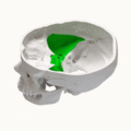
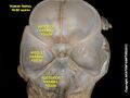
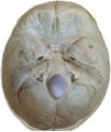
The middle cranial fossa is bounded anteriorly by the lesser wings of the sphenoid bone, posteriorly by the petrous ridges of the temporal bones, and laterally by the sphenoid and temporal bones. The floor of the middle cranial fossa is formed primarily by the sphenoid and temporal bones. It is divided into three main parts: the chiasmatic groove, the sella turcica, and the temporal fossa.
Chiasmatic Groove[edit]
The chiasmatic groove is a shallow sulcus that leads posteriorly to the optic canal, through which the optic nerve (CN II) and ophthalmic artery pass.
Sella Turcica[edit]
The sella turcica, a saddle-shaped depression in the body of the sphenoid bone, houses the pituitary gland. This vital structure is involved in the regulation of several hormones that control various bodily functions.
Temporal Fossa[edit]
The temporal fossa contains the temporal lobes of the brain and is crucial for the processing of auditory information and memory.
Clinical Significance[edit]
The middle cranial fossa is of significant clinical importance due to its contents and the conditions associated with it. Conditions such as pituitary adenomas, meningiomas, and cerebrospinal fluid leaks can manifest with symptoms related to the structures within the middle cranial fossa. Surgical approaches to the middle cranial fossa require intricate knowledge of its anatomy to avoid damage to the cranial nerves and brain tissue.
Surgical Approaches[edit]
Surgical approaches to the middle cranial fossa are complex and are typically performed to remove tumors, repair cerebrospinal fluid leaks, or address trigeminal neuralgia. These approaches must carefully navigate the dense concentration of nerves and blood vessels within this region.
See Also[edit]
This medical article is a stub. You can help WikiMD by expanding the page. |
-
Middle cranial fossa surgical anatomy
-
 Middle cranial fossa animation
 -
 Cranial endobasis of a 19-20 weeks foetus
 -
 Schädelbasis
 -
Base of skull
-
Base of skull
-
Middle cranial fossa


Medical Disclaimer: WikiMD is for informational purposes only and is not a substitute for professional medical advice. Content may be inaccurate or outdated and should not be used for diagnosis or treatment. Always consult your healthcare provider for medical decisions. Verify information with trusted sources such as CDC.gov and NIH.gov. By using this site, you agree that WikiMD is not liable for any outcomes related to its content. See full disclaimer.
Credits:Most images are courtesy of Wikimedia commons, and templates, categories Wikipedia, licensed under CC BY SA or similar.
Translate page: - East Asian
中文,
日本,
한국어,
South Asian
हिन्दी,
தமிழ்,
తెలుగు,
Urdu,
ಕನ್ನಡ,
Southeast Asian
Indonesian,
Vietnamese,
Thai,
မြန်မာဘာသာ,
বাংলা
European
español,
Deutsch,
français,
Greek,
português do Brasil,
polski,
română,
русский,
Nederlands,
norsk,
svenska,
suomi,
Italian
Middle Eastern & African
عربى,
Turkish,
Persian,
Hebrew,
Afrikaans,
isiZulu,
Kiswahili,
Other
Bulgarian,
Hungarian,
Czech,
Swedish,
മലയാളം,
मराठी,
ਪੰਜਾਬੀ,
ગુજરાતી,
Portuguese,
Ukrainian
