Middle cerebral artery
An article about the middle cerebral artery, its anatomy, function, and clinical significance
Anatomy[edit]
The middle cerebral artery (MCA) is one of the three major paired arteries that supply blood to the brain. It is a critical component of the cerebral circulation and is the most common site of stroke.
Origin[edit]
The MCA originates from the internal carotid artery and is considered a continuation of this artery. It is the largest branch of the internal carotid artery and supplies a significant portion of the lateral aspect of the brain.
Course[edit]
The MCA travels laterally into the Sylvian fissure, where it bifurcates into superior and inferior divisions. These divisions further branch into smaller arteries that supply the lateral surfaces of the frontal lobe, parietal lobe, and temporal lobe.
Branches[edit]
The branches of the MCA include:
- Lateral lenticulostriate arteries: These small, deep penetrating arteries supply the basal ganglia and the internal capsule.
- Cortical branches: These branches supply the lateral aspects of the cerebral cortex, including the primary motor and sensory areas.
Function[edit]
The MCA is responsible for supplying oxygenated blood to a large portion of the brain. It provides blood to areas involved in motor and sensory functions, language, and cognition. The regions supplied by the MCA are crucial for voluntary movement, sensory perception, and higher cognitive functions.
Clinical Significance[edit]
The MCA is the most common site of ischemic stroke. Occlusion of the MCA can lead to significant neurological deficits, depending on the location and extent of the blockage.
Stroke[edit]
A stroke in the territory of the MCA can result in:
- Contralateral hemiparesis: Weakness on the opposite side of the body.
- Contralateral sensory loss: Loss of sensation on the opposite side of the body.
- Aphasia: Difficulty with language, particularly if the dominant hemisphere is affected.
- Hemianopia: Loss of vision in half of the visual field.
Aneurysms[edit]
Aneurysms can occur in the MCA, leading to the risk of rupture and subarachnoid hemorrhage. These are often detected using imaging techniques such as CT scan or MRI.
Imaging[edit]
The MCA can be visualized using various imaging modalities, including:
- CT angiography: Provides detailed images of the blood vessels.
- Magnetic resonance angiography (MRA): Non-invasive imaging technique to visualize blood vessels.
Related pages[edit]
Middle_cerebral_artery[edit]
-
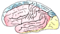 Gray's Anatomy plate 517 brain
 -
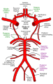 Circle of Willis en
 -
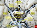 Circle of Willis 6
 -
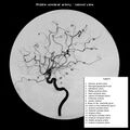 MCA angio lateral


Medical Disclaimer: WikiMD is for informational purposes only and is not a substitute for professional medical advice. Content may be inaccurate or outdated and should not be used for diagnosis or treatment. Always consult your healthcare provider for medical decisions. Verify information with trusted sources such as CDC.gov and NIH.gov. By using this site, you agree that WikiMD is not liable for any outcomes related to its content. See full disclaimer.
Credits:Most images are courtesy of Wikimedia commons, and templates, categories Wikipedia, licensed under CC BY SA or similar.
Translate page: - East Asian
中文,
日本,
한국어,
South Asian
हिन्दी,
தமிழ்,
తెలుగు,
Urdu,
ಕನ್ನಡ,
Southeast Asian
Indonesian,
Vietnamese,
Thai,
မြန်မာဘာသာ,
বাংলা
European
español,
Deutsch,
français,
Greek,
português do Brasil,
polski,
română,
русский,
Nederlands,
norsk,
svenska,
suomi,
Italian
Middle Eastern & African
عربى,
Turkish,
Persian,
Hebrew,
Afrikaans,
isiZulu,
Kiswahili,
Other
Bulgarian,
Hungarian,
Czech,
Swedish,
മലയാളം,
मराठी,
ਪੰਜਾਬੀ,
ગુજરાતી,
Portuguese,
Ukrainian
