Intercalated disc
Intercalated disc is a specialized structure found in the cardiac muscle tissue. It plays a crucial role in connecting adjacent cardiac muscle cells and facilitating the synchronized contraction of the heart muscle.
Structure[edit]
The intercalated disc is composed of three different types of cell junctions: desmosomes, gap junctions, and adherens junctions. These junctions allow for the rapid spread of electrical impulses between cardiac muscle cells, enabling the heart to contract as a single unit.
Desmosomes[edit]
Desmosomes are a type of cell junction that anchors adjacent cells together and resists mechanical stress. In the intercalated disc, desmosomes prevent cardiac muscle cells from being pulled apart during contraction.
Gap Junctions[edit]
Gap junctions are channels that allow for the direct passage of ions and small molecules between cells. In the intercalated disc, gap junctions facilitate the rapid spread of electrical impulses from one cardiac muscle cell to the next.
Adherens Junctions[edit]
Adherens junctions are a type of cell junction that connects the actin cytoskeleton of one cell to that of the neighboring cell. In the intercalated disc, adherens junctions help to coordinate the contraction of adjacent cardiac muscle cells.
Function[edit]
The primary function of the intercalated disc is to enable the heart to contract as a single unit. This is achieved through the rapid spread of electrical impulses from one cardiac muscle cell to the next, facilitated by the gap junctions in the intercalated disc.
In addition to this, the intercalated disc also plays a role in maintaining the structural integrity of the heart. The desmosomes in the intercalated disc prevent cardiac muscle cells from being pulled apart during contraction, while the adherens junctions help to coordinate the contraction of adjacent cells.
Clinical Significance[edit]
Abnormalities in the intercalated disc can lead to a variety of cardiac conditions. For example, mutations in the genes encoding for the proteins found in desmosomes can result in arrhythmogenic right ventricular cardiomyopathy (ARVC), a condition characterized by arrhythmias and heart failure.

This WikiMD article can only be edited by registered and verified editors. You can log in or register.
-
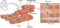
 Intercalated disc in cardiac muscle
 -
 Cardiac muscle showing intercalated discs
 -
 Histopathology of ruptured intercalated discs
 -
 Cardiac myofiber break-up with intercalated discs


Medical Disclaimer: WikiMD is for informational purposes only and is not a substitute for professional medical advice. Content may be inaccurate or outdated and should not be used for diagnosis or treatment. Always consult your healthcare provider for medical decisions. Verify information with trusted sources such as CDC.gov and NIH.gov. By using this site, you agree that WikiMD is not liable for any outcomes related to its content. See full disclaimer.
Credits:Most images are courtesy of Wikimedia commons, and templates, categories Wikipedia, licensed under CC BY SA or similar.
Translate page: - East Asian
中文,
日本,
한국어,
South Asian
हिन्दी,
தமிழ்,
తెలుగు,
Urdu,
ಕನ್ನಡ,
Southeast Asian
Indonesian,
Vietnamese,
Thai,
မြန်မာဘာသာ,
বাংলা
European
español,
Deutsch,
français,
Greek,
português do Brasil,
polski,
română,
русский,
Nederlands,
norsk,
svenska,
suomi,
Italian
Middle Eastern & African
عربى,
Turkish,
Persian,
Hebrew,
Afrikaans,
isiZulu,
Kiswahili,
Other
Bulgarian,
Hungarian,
Czech,
Swedish,
മലയാളം,
मराठी,
ਪੰਜਾਬੀ,
ગુજરાતી,
Portuguese,
Ukrainian
