In situ hybridization
In situ hybridization (ISH) is a type of hybridization that uses a labeled complementary DNA, RNA or modified nucleic acids strand (i.e., probe) to localize a specific DNA or RNA sequence in a portion or section of tissue (in situ), or, if the tissue is small enough (e.g., plant seeds, Drosophila embryos), in the entire tissue (whole mount ISH), in cells, and in circulating tumor cells (CTCs). This is distinct from immunohistochemistry, which usually localizes proteins in tissue sections.
Procedure[edit]
The procedure of in situ hybridization comprises the following steps:
- Fixation of tissue section or cells onto a glass slide
- Pre-hybridization treatment to increase accessibility of target DNA or RNA
- Hybridization of the probe to the DNA or RNA target
- Stringent washes to remove excess probe
- Detection of the hybridized probe
- Visualization of the target molecule
Applications[edit]
In situ hybridization is used in many areas of research and diagnosis including:
- Developmental biology
- Cancer and other disease diagnosis
- Genetics
- Evolutionary biology
Types of In Situ Hybridization[edit]
There are two types of in situ hybridization:
- Fluorescent in situ hybridization (FISH)
- Chromogenic in situ hybridization (CISH)
Fluorescent In Situ Hybridization (FISH)[edit]
Fluorescent in situ hybridization (FISH) is a molecular cytogenetic technique that uses fluorescent probes that bind to only those parts of the chromosome with a high degree of sequence complementarity.
Chromogenic In Situ Hybridization (CISH)[edit]
Chromogenic in situ hybridization (CISH) is a molecular technique that allows the visualization of specific genes or gene fragments in tissues.
See Also[edit]

This WikiMD article can only be edited by registered and verified editors. You can log in or register.
-
 RNA in situ hybridization in FFPE samples
 -
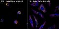 Multiplex ViewRNA FISH Assay in Jurkat and HeLa cells
 -
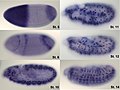 Hunchback in situ
 -
 HCR-FISH visualization of collagen expression in P. waltl


Medical Disclaimer: WikiMD is for informational purposes only and is not a substitute for professional medical advice. Content may be inaccurate or outdated and should not be used for diagnosis or treatment. Always consult your healthcare provider for medical decisions. Verify information with trusted sources such as CDC.gov and NIH.gov. By using this site, you agree that WikiMD is not liable for any outcomes related to its content. See full disclaimer.
Credits:Most images are courtesy of Wikimedia commons, and templates, categories Wikipedia, licensed under CC BY SA or similar.
Translate page: - East Asian
中文,
日本,
한국어,
South Asian
हिन्दी,
தமிழ்,
తెలుగు,
Urdu,
ಕನ್ನಡ,
Southeast Asian
Indonesian,
Vietnamese,
Thai,
မြန်မာဘာသာ,
বাংলা
European
español,
Deutsch,
français,
Greek,
português do Brasil,
polski,
română,
русский,
Nederlands,
norsk,
svenska,
suomi,
Italian
Middle Eastern & African
عربى,
Turkish,
Persian,
Hebrew,
Afrikaans,
isiZulu,
Kiswahili,
Other
Bulgarian,
Hungarian,
Czech,
Swedish,
മലയാളം,
मराठी,
ਪੰਜਾਬੀ,
ગુજરાતી,
Portuguese,
Ukrainian
