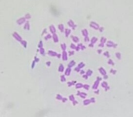
G banding
Technique for staining chromosomes
G banding (or Giemsa banding) is a technique used in cytogenetics to produce a visible karyotype by staining condensed chromosomes. It is one of the most common methods for identifying chromosomal abnormalities and for chromosome mapping.
Principle[edit]
G banding involves treating chromosomes with a proteolytic enzyme, such as trypsin, followed by staining with Giemsa stain. The Giemsa stain binds to the phosphate groups of DNA, but it stains regions rich in adenine (A) and thymine (T) more intensely. This results in a pattern of light and dark bands, known as G bands, along the length of each chromosome.
Procedure[edit]
The process of G banding involves several steps:
- Cell Culture: Cells are cultured in vitro to increase their number. Lymphocytes from blood samples are commonly used.
- Cell Harvesting: Cells are arrested in metaphase using a mitotic inhibitor such as colchicine.
- Hypotonic Treatment: Cells are treated with a hypotonic solution to swell them, making the chromosomes more spread out.
- Fixation: Cells are fixed using a mixture of methanol and acetic acid.
- Slide Preparation: The fixed cells are dropped onto a microscope slide, where they burst open, spreading the chromosomes.
- Enzyme Treatment: The slide is treated with trypsin to partially digest the proteins associated with the chromosomes.
- Staining: The slide is stained with Giemsa stain, which binds to the DNA and produces the characteristic banding pattern.
Applications[edit]
G banding is used in various applications, including:
- Karyotyping: To identify and evaluate the size, shape, and number of chromosomes in a sample of body cells.
- Diagnosis of Genetic Disorders: To detect chromosomal abnormalities such as Down syndrome, Klinefelter syndrome, and Turner syndrome.
- Cancer Cytogenetics: To identify chromosomal changes in cancer cells, which can help in diagnosis and treatment planning.
- Prenatal Diagnosis: To assess the chromosomal health of a fetus using amniocentesis or chorionic villus sampling.
Advantages and Limitations[edit]
Advantages[edit]
- High Resolution: G banding provides a high-resolution view of chromosomes, allowing for detailed analysis.
- Reproducibility: The technique is well-established and reproducible across different laboratories.
Limitations[edit]
- Resolution Limit: G banding cannot detect very small chromosomal changes, such as microdeletions or microduplications.
- Requires Dividing Cells: The technique requires cells to be in metaphase, which limits its use to dividing cells.
Images[edit]


Related pages[edit]
Ad. Transform your life with W8MD's Budget GLP-1 injections from $75


W8MD offers a medical weight loss program to lose weight in Philadelphia. Our physician-supervised medical weight loss provides:
- Weight loss injections in NYC (generic and brand names):
- Zepbound / Mounjaro, Wegovy / Ozempic, Saxenda
- Most insurances accepted or discounted self-pay rates. We will obtain insurance prior authorizations if needed.
- Generic GLP1 weight loss injections from $75 for the starting dose.
- Also offer prescription weight loss medications including Phentermine, Qsymia, Diethylpropion, Contrave etc.
NYC weight loss doctor appointmentsNYC weight loss doctor appointments
Start your NYC weight loss journey today at our NYC medical weight loss and Philadelphia medical weight loss clinics.
- Call 718-946-5500 to lose weight in NYC or for medical weight loss in Philadelphia 215-676-2334.
- Tags:NYC medical weight loss, Philadelphia lose weight Zepbound NYC, Budget GLP1 weight loss injections, Wegovy Philadelphia, Wegovy NYC, Philadelphia medical weight loss, Brookly weight loss and Wegovy NYC
|
WikiMD's Wellness Encyclopedia |
| Let Food Be Thy Medicine Medicine Thy Food - Hippocrates |
Medical Disclaimer: WikiMD is not a substitute for professional medical advice. The information on WikiMD is provided as an information resource only, may be incorrect, outdated or misleading, and is not to be used or relied on for any diagnostic or treatment purposes. Please consult your health care provider before making any healthcare decisions or for guidance about a specific medical condition. WikiMD expressly disclaims responsibility, and shall have no liability, for any damages, loss, injury, or liability whatsoever suffered as a result of your reliance on the information contained in this site. By visiting this site you agree to the foregoing terms and conditions, which may from time to time be changed or supplemented by WikiMD. If you do not agree to the foregoing terms and conditions, you should not enter or use this site. See full disclaimer.
Credits:Most images are courtesy of Wikimedia commons, and templates, categories Wikipedia, licensed under CC BY SA or similar.
Translate this page: - East Asian
中文,
日本,
한국어,
South Asian
हिन्दी,
தமிழ்,
తెలుగు,
Urdu,
ಕನ್ನಡ,
Southeast Asian
Indonesian,
Vietnamese,
Thai,
မြန်မာဘာသာ,
বাংলা
European
español,
Deutsch,
français,
Greek,
português do Brasil,
polski,
română,
русский,
Nederlands,
norsk,
svenska,
suomi,
Italian
Middle Eastern & African
عربى,
Turkish,
Persian,
Hebrew,
Afrikaans,
isiZulu,
Kiswahili,
Other
Bulgarian,
Hungarian,
Czech,
Swedish,
മലയാളം,
मराठी,
ਪੰਜਾਬੀ,
ગુજરાતી,
Portuguese,
Ukrainian