Foreign-body giant cell
Foreign-body giant cell (FBGC) is a type of giant cell that forms when the immune system attempts to isolate and attack foreign materials in the body that are too large to be engulfed by individual macrophages. These cells are characterized by their large size, with multiple nuclei arranged in a ring or scattered throughout the cell. FBGCs are commonly observed in chronic inflammation cases, particularly in response to non-biodegradable materials, and play a significant role in the foreign body reaction (FBR).
Formation[edit]
FBGCs arise from the fusion of monocytes or macrophages, which are types of white blood cells that play a crucial role in the body's immune response. When these cells encounter a foreign material that they cannot phagocytize or break down due to its size or nature, they release cytokines and growth factors that promote the fusion of adjacent macrophages, leading to the formation of a giant cell with multiple nuclei.
Function[edit]
The primary function of FBGCs is to attempt to degrade or isolate foreign materials that are recognized as non-self by the immune system. These materials can include medical implants, sutures, and other non-biodegradable objects introduced into the body. Despite their efforts, FBGCs are often unable to completely degrade the foreign material, leading to a persistent foreign body reaction. This reaction can result in the formation of a fibrous capsule around the material, effectively isolating it from the surrounding tissue.
Clinical Significance[edit]
FBGCs are of clinical significance because they are indicative of a chronic inflammatory response to foreign materials in the body. Their presence can lead to complications in wound healing and the integration of medical implants. Understanding the formation and function of FBGCs is crucial for the development of biocompatible materials that minimize the foreign body reaction and improve the outcomes of medical implants and other procedures involving foreign materials.
Histopathology[edit]
Histologically, FBGCs can be identified by their large size, multiple nuclei, and the presence of a surrounding inflammatory infiltrate. Special staining techniques, such as hematoxylin and eosin (H&E) staining, can be used to visualize these cells in tissue samples.
Management[edit]
Management of conditions involving FBGCs typically focuses on removing the foreign material, if possible, and addressing the underlying inflammation. In cases where the foreign material cannot be removed, treatment may involve the use of anti-inflammatory medications to reduce the immune response.
See Also[edit]
This medical article is a stub. You can help WikiMD by expanding the page. |
-
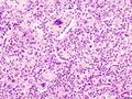 Aspiration pneumonia micrograph
 -
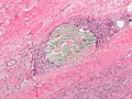 Suture micrograph
 -
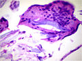 Foreign body engulfed by a giant cell
 -
 Silicone breast implant
.jpg)


Medical Disclaimer: WikiMD is for informational purposes only and is not a substitute for professional medical advice. Content may be inaccurate or outdated and should not be used for diagnosis or treatment. Always consult your healthcare provider for medical decisions. Verify information with trusted sources such as CDC.gov and NIH.gov. By using this site, you agree that WikiMD is not liable for any outcomes related to its content. See full disclaimer.
Credits:Most images are courtesy of Wikimedia commons, and templates, categories Wikipedia, licensed under CC BY SA or similar.
Translate page: - East Asian
中文,
日本,
한국어,
South Asian
हिन्दी,
தமிழ்,
తెలుగు,
Urdu,
ಕನ್ನಡ,
Southeast Asian
Indonesian,
Vietnamese,
Thai,
မြန်မာဘာသာ,
বাংলা
European
español,
Deutsch,
français,
Greek,
português do Brasil,
polski,
română,
русский,
Nederlands,
norsk,
svenska,
suomi,
Italian
Middle Eastern & African
عربى,
Turkish,
Persian,
Hebrew,
Afrikaans,
isiZulu,
Kiswahili,
Other
Bulgarian,
Hungarian,
Czech,
Swedish,
മലയാളം,
मराठी,
ਪੰਜਾਬੀ,
ગુજરાતી,
Portuguese,
Ukrainian
