File:XiiSchematic.jpg
From WikiMD's medical encyclopedia
No higher resolution available.
XiiSchematic.jpg (692 × 356 pixels, file size: 104 KB, MIME type: image/jpeg)
Summary
| Description |
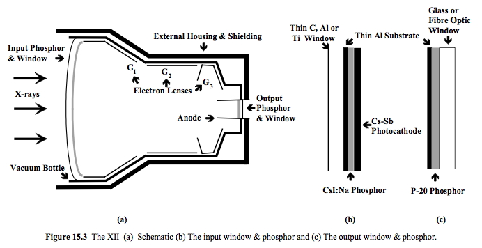
English: The X-ray image intensifier (a), with close-ups of the phosphor/photocathode sandwich (b) and the output phosphor (c). |
| Date | |
| Source | Own work |
| Author | Kieranmaher |
Reproduced with permission from Heggie JCP, Liddell NA & Maher KP, 2001, Applied Imaging Technology, 4th Edition
Licensing
| I, the copyright holder of this work, release this work into the public domain. This applies worldwide. In some countries this may not be legally possible; if so: I grant anyone the right to use this work for any purpose, without any conditions, unless such conditions are required by law. |

|
This physics image could be re-created using vector graphics as an SVG file. This has several advantages; see Commons:Media for cleanup for more information. If an SVG form of this image is available, please upload it and afterwards replace this template with
{{vector version available|new image name}}.It is recommended to name the SVG file “XiiSchematic.svg”—then the template Vector version available (or Vva) does not need the new image name parameter. |


.
File history
Click on a date/time to view the file as it appeared at that time.
| Date/Time | Thumbnail | Dimensions | User | Comment | |
|---|---|---|---|---|---|
| current | 07:37, 23 April 2013 | | 692 × 356 (104 KB) | Bidgee | If you want to request deletion, please create a mass deletion request |
File usage
The following page uses this file: