File:CT colonography of a rectal mass.jpg
From WikiMD's medical encyclopedia
No higher resolution available.
CT_colonography_of_a_rectal_mass.jpg (665 × 305 pixels, file size: 56 KB, MIME type: image/jpeg)
Summary
| Description |
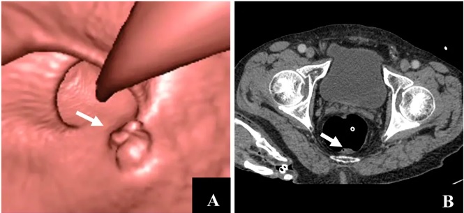
English: CT colonography of a rectal mass. Left image is a volume rendering and right image is a thin slice. It also shows the rectal tube used for insufflation of gas to distend the colon.
Original caption: Differentiation from stool and colonic neoplasia by dual-energy CT. Male, 88 years of age. (A,B) denote colonography and CT axial plane image revealed irregular upheaval masses in the posterior wall of the rectum (white arrow). It is difficult to differentiate tumors from stool. Dual-energy contrast-enhanced CT (axial plane) revealed no enhancement of masses, iodine value = −1.4 mg/ml (white arrow), suggesting no blood supply to the masses, which was validated as stool by colonography. |
| Date | |
| Source |
(2018). "Accuracy of Combined Computed Tomography Colonography and Dual Energy Iiodine Map Imaging for Detecting Colorectal masses using High-pitch Dual-source CT". Scientific Reports 8 (1). DOI:10.1038/s41598-018-22188-x. ISSN 2045-2322.
|
| Author | Kai Sun, Ruijuan Han, Yang Han, Xuesen Shi, Jiang Hu & Bin lu |
Licensing
This file is licensed under the Creative Commons Attribution 4.0 International license.
- You are free:
- to share – to copy, distribute and transmit the work
- to remix – to adapt the work
- Under the following conditions:
- attribution – You must give appropriate credit, provide a link to the license, and indicate if changes were made. You may do so in any reasonable manner, but not in any way that suggests the licensor endorses you or your use.
File history
Click on a date/time to view the file as it appeared at that time.
| Date/Time | Thumbnail | Dimensions | User | Comment | |
|---|---|---|---|---|---|
| current | 08:28, 26 July 2019 | | 665 × 305 (56 KB) | Mikael Häggström | User created page with UploadWizard |
File usage
The following page uses this file: