File:2B8T.png
From WikiMD's medical encyclopedia

Size of this preview: 800 × 600 pixels. Other resolutions: 320 × 240 pixels | 640 × 480 pixels | 1,024 × 768 pixels | 1,280 × 960 pixels | 1,440 × 1,080 pixels.
Original file (1,440 × 1,080 pixels, file size: 661 KB, MIME type: image/png)
Summary
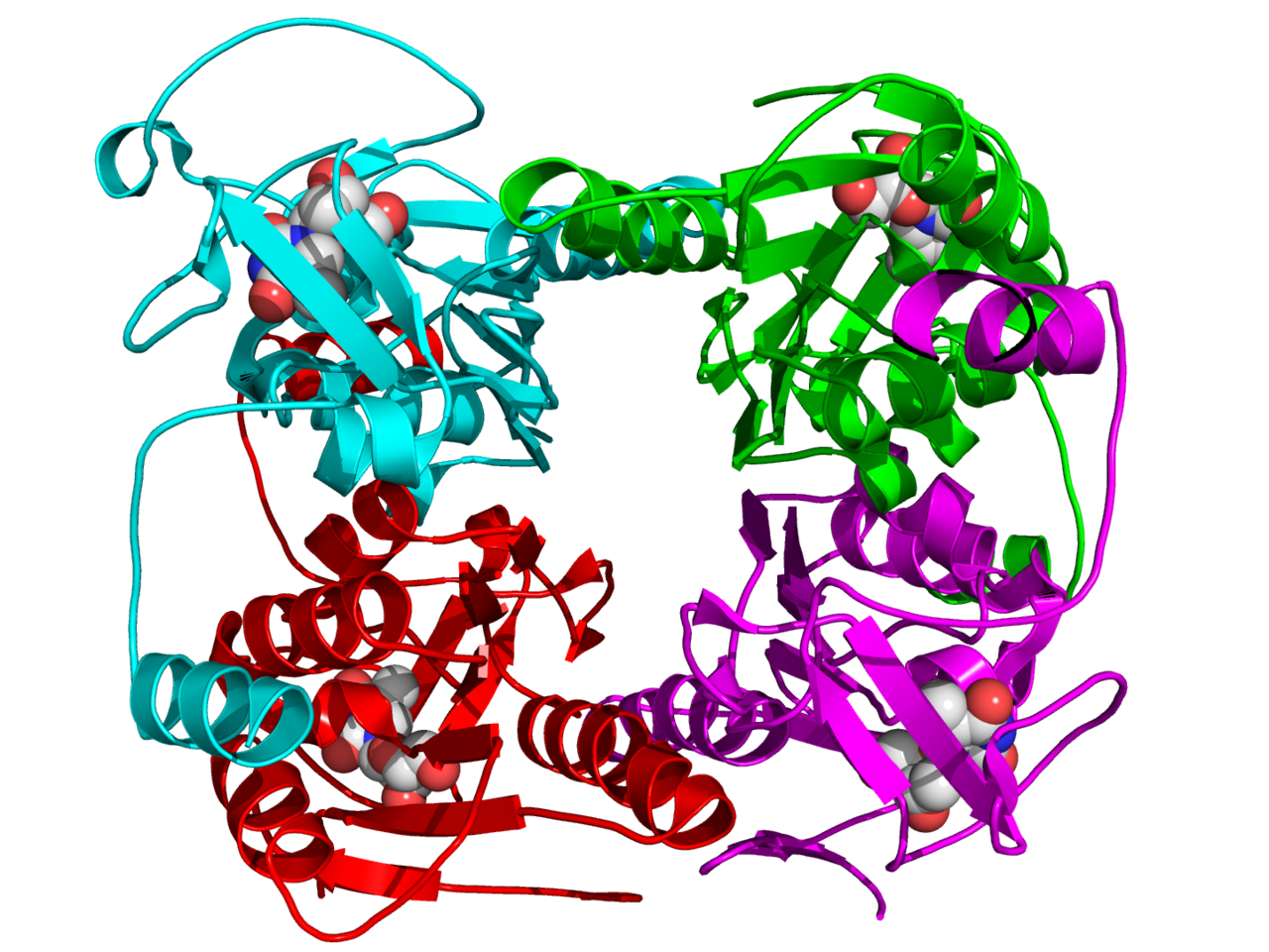
| Description | Crystal structure of a tetramer of thymidine kinase from Ureaplasma urealyticum (where the monomers are color cyan, green, red, and magenta respectively) in complex with thymidine (space-filling model, carbon = white, oxygen = red, nitrogen = blue).[1] |
| Date | |
| Source | Own work |
| Author | Boghog2 |
References
- ↑ PDB: 2B8T; Kosinska U, Carnrot C, Eriksson S, Wang L, Eklund H (December 2005). "Structure of the substrate complex of thymidine kinase from Ureaplasma urealyticum and investigations of possible drug targets for the enzyme". FEBS J. 272 (24): 6365–72. DOI:10.1111/j.1742-4658.2005.05030.x. PMID 16336273.
Licensing
| I, the copyright holder of this work, release this work into the public domain. This applies worldwide. In some countries this may not be legally possible; if so: I grant anyone the right to use this work for any purpose, without any conditions, unless such conditions are required by law. |
File history
Click on a date/time to view the file as it appeared at that time.
| Date/Time | Thumbnail | Dimensions | User | Comment | |
|---|---|---|---|---|---|
| current | 10:52, 22 November 2008 | | 1,440 × 1,080 (661 KB) | Boghog | {{Information |Description=Crystal structure of a tetramer of thymidine kinase from ''U. urealyticum'' (where the monomers are color cyan, green, red, and magenta respectively) in complex with thymidine (space-filling model, carbon = white, ox |
File usage
The following page uses this file: