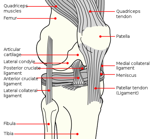
Anterior cruciate ligament

The anterior cruciate ligament (ACL) is one of the major ligaments in the human knee, playing a pivotal role in knee stability and movement. Its primary function is to prevent excessive forward movement of the tibia in relation to the femur and provide rotational stability to the knee. Injuries to the ACL are common, especially in athletes, and can significantly impact an individual's mobility and quality of life.
Anatomy[edit]
The ACL is one of the four primary ligaments in the knee. It originates from the posterior aspect of the medial side of the lateral femoral condyle and attaches to the anterior intercondylar area of the tibia.
Key structures associated with the ACL include:
- PCL (Posterior Cruciate Ligament): Another major ligament in the knee, located just behind the ACL.
- Menisci: Fibrocartilage pads that lie between the femur and tibia.
- Synovial Membrane: Surrounds the ACL and produces synovial fluid for joint lubrication.
Function[edit]
The ACL serves several crucial functions in the knee:
- Rotational Stability: Prevents excessive rotation of the tibia.
- Anteroposterior Stability: Stops the tibia from sliding too far forward under the femur.
- Proprioception: Contains nerve endings that help in sensing the position of the joint.
Injuries[edit]
ACL injuries are among the most common knee injuries, especially in sports:
- Mechanism of Injury: Typically, a non-contact pivoting injury. Direct contact, however, can also result in an ACL tear.
- Symptoms: Often, there's an audible "pop" during injury, followed by swelling, pain, and instability.
- Diagnosis: Typically through clinical examination and confirmed with MRI.
- Treatment:
- Conservative: Physical therapy and strengthening exercises.
- Surgical: ACL reconstruction, usually using a graft from another ligament or tendon in the body.
Rehabilitation and Recovery[edit]
Following an ACL injury or surgery, rehabilitation is crucial:
- Physical Therapy: Focuses on restoring movement, strength, and function to the knee.
- Bracing: Some patients may benefit from wearing a knee brace during recovery.
- Return to Activity: Gradual and typically under the guidance of a healthcare professional.
Prevention[edit]
Prevention of ACL injuries includes:
- Strength Training: Especially of the hamstrings and quadriceps.
- Plyometric Drills: Jumping exercises to improve agility and muscular response.
- Proper Technique: Educating athletes on proper techniques, especially in sports like soccer, basketball, and skiing.
- Bracing: For some athletes, especially those at higher risk or with a history of ACL injury.
| Joints and ligaments of the human leg | ||||||||||||||
|---|---|---|---|---|---|---|---|---|---|---|---|---|---|---|
|
Ad. Transform your life with W8MD's Budget GLP-1 injections from $75


W8MD offers a medical weight loss program to lose weight in Philadelphia. Our physician-supervised medical weight loss provides:
- Weight loss injections in NYC (generic and brand names):
- Zepbound / Mounjaro, Wegovy / Ozempic, Saxenda
- Most insurances accepted or discounted self-pay rates. We will obtain insurance prior authorizations if needed.
- Generic GLP1 weight loss injections from $75 for the starting dose.
- Also offer prescription weight loss medications including Phentermine, Qsymia, Diethylpropion, Contrave etc.
NYC weight loss doctor appointmentsNYC weight loss doctor appointments
Start your NYC weight loss journey today at our NYC medical weight loss and Philadelphia medical weight loss clinics.
- Call 718-946-5500 to lose weight in NYC or for medical weight loss in Philadelphia 215-676-2334.
- Tags:NYC medical weight loss, Philadelphia lose weight Zepbound NYC, Budget GLP1 weight loss injections, Wegovy Philadelphia, Wegovy NYC, Philadelphia medical weight loss, Brookly weight loss and Wegovy NYC
|
WikiMD's Wellness Encyclopedia |
| Let Food Be Thy Medicine Medicine Thy Food - Hippocrates |
Medical Disclaimer: WikiMD is not a substitute for professional medical advice. The information on WikiMD is provided as an information resource only, may be incorrect, outdated or misleading, and is not to be used or relied on for any diagnostic or treatment purposes. Please consult your health care provider before making any healthcare decisions or for guidance about a specific medical condition. WikiMD expressly disclaims responsibility, and shall have no liability, for any damages, loss, injury, or liability whatsoever suffered as a result of your reliance on the information contained in this site. By visiting this site you agree to the foregoing terms and conditions, which may from time to time be changed or supplemented by WikiMD. If you do not agree to the foregoing terms and conditions, you should not enter or use this site. See full disclaimer.
Credits:Most images are courtesy of Wikimedia commons, and templates, categories Wikipedia, licensed under CC BY SA or similar.
Translate this page: - East Asian
中文,
日本,
한국어,
South Asian
हिन्दी,
தமிழ்,
తెలుగు,
Urdu,
ಕನ್ನಡ,
Southeast Asian
Indonesian,
Vietnamese,
Thai,
မြန်မာဘာသာ,
বাংলা
European
español,
Deutsch,
français,
Greek,
português do Brasil,
polski,
română,
русский,
Nederlands,
norsk,
svenska,
suomi,
Italian
Middle Eastern & African
عربى,
Turkish,
Persian,
Hebrew,
Afrikaans,
isiZulu,
Kiswahili,
Other
Bulgarian,
Hungarian,
Czech,
Swedish,
മലയാളം,
मराठी,
ਪੰਜਾਬੀ,
ગુજરાતી,
Portuguese,
Ukrainian
