Renal ultrasonography
Renal ultrasonography is a common diagnostic procedure used in the field of nephrology to assess the structure and function of the kidney. It is a non-invasive method that uses ultrasound waves to create images of the kidneys and surrounding structures.
Overview[edit]
Renal ultrasonography is a type of medical imaging that uses high-frequency sound waves to produce images of the kidneys. The procedure is performed by a radiologist or a sonographer who uses an ultrasound machine and a transducer to send sound waves into the body. These sound waves bounce off the kidneys and other structures, and their echoes are picked up by the transducer and converted into images.
Indications[edit]
Renal ultrasonography is used for a variety of reasons. It can be used to evaluate the size, shape, and position of the kidneys, to detect abnormalities such as kidney stones, cysts, and tumors, and to assess blood flow to the kidneys. It is also used to guide procedures such as kidney biopsy and nephrostomy.
Procedure[edit]
During a renal ultrasonography, the patient lies on an examination table and a clear gel is applied to the skin over the kidneys. The sonographer moves the transducer over the area to obtain images of the kidneys and surrounding structures. The procedure is painless and typically takes about 30 minutes to complete.
Risks and Complications[edit]
Renal ultrasonography is considered a safe procedure with no known risks or complications. It does not use radiation, making it a good choice for patients who are pregnant or have certain medical conditions that make other types of imaging risky.
See Also[edit]

-
 Doppler ultrasound of systolic velocity (Vs), diastolic velocity (Vd), acceleration time (AoAT), systolic acceleration (Ao Accel) and resistive index (RI) of normal kidney
 -
 Normal adult kidney
 -
 Normal pediatric kidney
 -
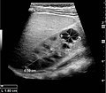
 Measures of the kidney
 -
 Simple cyst with posterior enhancement
 -
 Complex cyst with thickened walls and membranes in the lower pole
 -
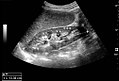
 Advanced polycystic kidney disease with multiple cysts
 -
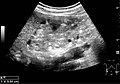
 Cortical solid mass of renal cell carcinoma
 -
 Renal cell carcinoma with both cystic and solid components
 -
 Ultrasonography of renal lymphoma
 -
 Ultrasonography of angiomyolipoma
 -
 Ultrasonography of multiple angiomyolipomas in tuberous sclerosis
 -
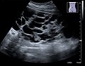
 Ultrasonography of hydronephrosis due to ureteropelvic junction obstruction
,_diastolic_velocity_(Vd),_acceleration_time_(AoAT),_systolic_acceleration_(Ao_Accel)_and_resistive_index_(RI)_of_normal_kidney.jpg)


Ad. Transform your life with W8MD's
GLP-1 weight loss injections special from $29.99 with insurance
|
WikiMD Medical Encyclopedia |
Medical Disclaimer: WikiMD is for informational purposes only and is not a substitute for professional medical advice. Content may be inaccurate or outdated and should not be used for diagnosis or treatment. Always consult your healthcare provider for medical decisions. Verify information with trusted sources such as CDC.gov and NIH.gov. By using this site, you agree that WikiMD is not liable for any outcomes related to its content. See full disclaimer.
Credits:Most images are courtesy of Wikimedia commons, and templates, categories Wikipedia, licensed under CC BY SA or similar.
Translate this page: - East Asian
中文,
日本,
한국어,
South Asian
हिन्दी,
தமிழ்,
తెలుగు,
Urdu,
ಕನ್ನಡ,
Southeast Asian
Indonesian,
Vietnamese,
Thai,
မြန်မာဘာသာ,
বাংলা
European
español,
Deutsch,
français,
Greek,
português do Brasil,
polski,
română,
русский,
Nederlands,
norsk,
svenska,
suomi,
Italian
Middle Eastern & African
عربى,
Turkish,
Persian,
Hebrew,
Afrikaans,
isiZulu,
Kiswahili,
Other
Bulgarian,
Hungarian,
Czech,
Swedish,
മലയാളം,
मराठी,
ਪੰਜਾਬੀ,
ગુજરાતી,
Portuguese,
Ukrainian
