Tinea nigra: Difference between revisions
CSV import |
CSV import |
||
| Line 1: | Line 1: | ||
[[file:Tinea_nigra_new_image.jpg|thumb|Tinea nigra new image]] '''Tinea nigra''' is a superficial fungal infection of the skin caused by the dematiaceous (darkly pigmented) fungus ''[[Hortaea werneckii]]''. This condition is characterized by brown to black, non-scaly patches on the palms of the hands and the soles of the feet. It is a relatively rare infection and is most commonly found in tropical and subtropical regions. | {{SI}} | ||
{{Infobox medical condition | |||
| name = Tinea nigra | |||
| image = [[File:Hortaea-werneckii-fungus--causes-tinea-nigra.jpg|alt=Hortaea werneckii fungus causes tinea nigra]] | |||
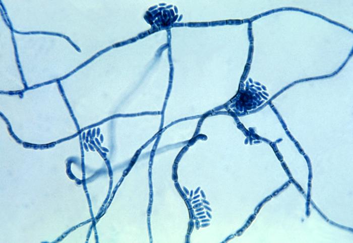
| caption = ''[[Hortaea werneckii]]'' fungus causes tinea nigra | |||
| field = [[Dermatology]] | |||
| synonyms = [[Keratomycosis nigricans palmaris]] | |||
| symptoms = [[Asymptomatic]] dark brown or black patches on [[palms]] or [[soles]] | |||
| complications = None | |||
| onset = Any age, more common in [[children]] and [[young adults]] | |||
| duration = Chronic if untreated | |||
| causes = ''[[Hortaea werneckii]]'' | |||
| risks = [[Warm]] and [[humid]] climates | |||
| diagnosis = [[Clinical diagnosis]], [[KOH test]], [[skin biopsy]] | |||
| differential = [[Melanoma]], [[nevus]], [[post-inflammatory hyperpigmentation]] | |||
| prevention = Avoidance of [[contaminated soil]] or [[water]] | |||
| treatment = [[Topical antifungal]]s such as [[miconazole]], [[clotrimazole]] | |||
| prognosis = Excellent with treatment | |||
| frequency = Rare | |||
}} | |||
[[file:Tinea_nigra_new_image.jpg|left|thumb|Tinea nigra new image]] '''Tinea nigra''' is a superficial fungal infection of the skin caused by the dematiaceous (darkly pigmented) fungus ''[[Hortaea werneckii]]''. This condition is characterized by brown to black, non-scaly patches on the palms of the hands and the soles of the feet. It is a relatively rare infection and is most commonly found in tropical and subtropical regions. | |||
==Presentation== | ==Presentation== | ||
The primary symptom of tinea nigra is the appearance of dark patches on the skin. These patches are usually asymptomatic, meaning they do not cause pain or itching. The lesions are typically well-defined, flat, and can vary in color from light brown to black. They are most commonly found on the palms and soles but can occasionally appear on other parts of the body. | The primary symptom of tinea nigra is the appearance of dark patches on the skin. These patches are usually asymptomatic, meaning they do not cause pain or itching. The lesions are typically well-defined, flat, and can vary in color from light brown to black. They are most commonly found on the palms and soles but can occasionally appear on other parts of the body. | ||
==Pathogenesis== | ==Pathogenesis== | ||
Tinea nigra is caused by the fungus ''[[Hortaea werneckii]]'', which is a type of [[dematiaceous fungi]]. The fungus produces melanin, which gives the lesions their characteristic dark color. The infection is usually acquired through direct contact with contaminated soil, wood, or decaying vegetation. The fungus thrives in warm, humid environments, which is why the infection is more prevalent in tropical and subtropical regions. | Tinea nigra is caused by the fungus ''[[Hortaea werneckii]]'', which is a type of [[dematiaceous fungi]]. The fungus produces melanin, which gives the lesions their characteristic dark color. The infection is usually acquired through direct contact with contaminated soil, wood, or decaying vegetation. The fungus thrives in warm, humid environments, which is why the infection is more prevalent in tropical and subtropical regions. | ||
==Diagnosis== | ==Diagnosis== | ||
Diagnosis of tinea nigra is typically made through clinical examination and confirmed by laboratory tests. A skin scraping from the affected area can be examined under a microscope after being treated with potassium hydroxide (KOH). The presence of darkly pigmented, septate hyphae and yeast-like cells is indicative of ''[[Hortaea werneckii]]''. Culture and histopathological examination can also be used to confirm the diagnosis. | Diagnosis of tinea nigra is typically made through clinical examination and confirmed by laboratory tests. A skin scraping from the affected area can be examined under a microscope after being treated with potassium hydroxide (KOH). The presence of darkly pigmented, septate hyphae and yeast-like cells is indicative of ''[[Hortaea werneckii]]''. Culture and histopathological examination can also be used to confirm the diagnosis. | ||
==Treatment== | ==Treatment== | ||
Treatment for tinea nigra is usually straightforward and involves the use of topical antifungal agents. Commonly used medications include [[miconazole]], [[clotrimazole]], and [[terbinafine]]. These treatments are typically applied to the affected area once or twice daily for several weeks. In some cases, keratolytic agents such as salicylic acid may be used to help remove the outer layer of skin and enhance the effectiveness of the antifungal treatment. | Treatment for tinea nigra is usually straightforward and involves the use of topical antifungal agents. Commonly used medications include [[miconazole]], [[clotrimazole]], and [[terbinafine]]. These treatments are typically applied to the affected area once or twice daily for several weeks. In some cases, keratolytic agents such as salicylic acid may be used to help remove the outer layer of skin and enhance the effectiveness of the antifungal treatment. | ||
==Prevention== | ==Prevention== | ||
Preventing tinea nigra involves minimizing exposure to the environmental sources of the fungus. This includes avoiding direct contact with contaminated soil, wood, and vegetation, especially in tropical and subtropical regions. Maintaining good personal hygiene and keeping the skin dry can also help reduce the risk of infection. | Preventing tinea nigra involves minimizing exposure to the environmental sources of the fungus. This includes avoiding direct contact with contaminated soil, wood, and vegetation, especially in tropical and subtropical regions. Maintaining good personal hygiene and keeping the skin dry can also help reduce the risk of infection. | ||
==See also== | |||
== | |||
* [[Dermatophyte]] | * [[Dermatophyte]] | ||
* [[Fungal infection]] | * [[Fungal infection]] | ||
| Line 23: | Line 37: | ||
* [[Hortaea werneckii]] | * [[Hortaea werneckii]] | ||
[[Category:Fungal diseases]] | [[Category:Fungal diseases]] | ||
[[Category:Skin conditions resulting from fungi]] | [[Category:Skin conditions resulting from fungi]] | ||
[[Category:Mycosis-related cutaneous conditions]] | [[Category:Mycosis-related cutaneous conditions]] | ||
[[Category:Infectious diseases]] | [[Category:Infectious diseases]] | ||
{{Infectious-disease-stub}} | {{Infectious-disease-stub}} | ||
Latest revision as of 15:34, 8 April 2025

Editor-In-Chief: Prab R Tumpati, MD
Obesity, Sleep & Internal medicine
Founder, WikiMD Wellnesspedia &
W8MD medical weight loss NYC and sleep center NYC
| Tinea nigra | |
|---|---|

| |
| Synonyms | Keratomycosis nigricans palmaris |
| Pronounce | N/A |
| Specialty | N/A |
| Symptoms | Asymptomatic dark brown or black patches on palms or soles |
| Complications | None |
| Onset | Any age, more common in children and young adults |
| Duration | Chronic if untreated |
| Types | N/A |
| Causes | Hortaea werneckii |
| Risks | Warm and humid climates |
| Diagnosis | Clinical diagnosis, KOH test, skin biopsy |
| Differential diagnosis | Melanoma, nevus, post-inflammatory hyperpigmentation |
| Prevention | Avoidance of contaminated soil or water |
| Treatment | Topical antifungals such as miconazole, clotrimazole |
| Medication | N/A |
| Prognosis | Excellent with treatment |
| Frequency | Rare |
| Deaths | N/A |

Tinea nigra is a superficial fungal infection of the skin caused by the dematiaceous (darkly pigmented) fungus Hortaea werneckii. This condition is characterized by brown to black, non-scaly patches on the palms of the hands and the soles of the feet. It is a relatively rare infection and is most commonly found in tropical and subtropical regions.
Presentation[edit]
The primary symptom of tinea nigra is the appearance of dark patches on the skin. These patches are usually asymptomatic, meaning they do not cause pain or itching. The lesions are typically well-defined, flat, and can vary in color from light brown to black. They are most commonly found on the palms and soles but can occasionally appear on other parts of the body.
Pathogenesis[edit]
Tinea nigra is caused by the fungus Hortaea werneckii, which is a type of dematiaceous fungi. The fungus produces melanin, which gives the lesions their characteristic dark color. The infection is usually acquired through direct contact with contaminated soil, wood, or decaying vegetation. The fungus thrives in warm, humid environments, which is why the infection is more prevalent in tropical and subtropical regions.
Diagnosis[edit]
Diagnosis of tinea nigra is typically made through clinical examination and confirmed by laboratory tests. A skin scraping from the affected area can be examined under a microscope after being treated with potassium hydroxide (KOH). The presence of darkly pigmented, septate hyphae and yeast-like cells is indicative of Hortaea werneckii. Culture and histopathological examination can also be used to confirm the diagnosis.
Treatment[edit]
Treatment for tinea nigra is usually straightforward and involves the use of topical antifungal agents. Commonly used medications include miconazole, clotrimazole, and terbinafine. These treatments are typically applied to the affected area once or twice daily for several weeks. In some cases, keratolytic agents such as salicylic acid may be used to help remove the outer layer of skin and enhance the effectiveness of the antifungal treatment.
Prevention[edit]
Preventing tinea nigra involves minimizing exposure to the environmental sources of the fungus. This includes avoiding direct contact with contaminated soil, wood, and vegetation, especially in tropical and subtropical regions. Maintaining good personal hygiene and keeping the skin dry can also help reduce the risk of infection.
See also[edit]

This article is a infectious disease stub. You can help WikiMD by expanding it!