Pyknosis: Difference between revisions
CSV import |
CSV import |
||
| Line 41: | Line 41: | ||
[[Category:Cellular processes]] | [[Category:Cellular processes]] | ||
{{medicine-stub}} | {{medicine-stub}} | ||
== Pyknosis == | |||
<gallery> | |||
File:Apoptosis.png|Apoptosis | |||
File:4_Bd_obs_4_680x512px.tif|4 Bd obs 4 680x512px | |||
File:nuclear_changes.jpg|Nuclear changes | |||
File:Histopathology_of_a_pheochromocytoma_with_coagulative_necrosis,_with_immunostaining.jpg|Histopathology of a pheochromocytoma with coagulative necrosis, with immunostaining | |||
File:Apoptotic_DNA_Laddering.png|Apoptotic DNA Laddering | |||
</gallery> | |||
Latest revision as of 21:05, 23 February 2025

Pyknosis is a term used in cell biology to describe the irreversible condensation of chromatin in the nucleus of a cell undergoing apoptosis or necrosis. This process is a hallmark of cell death and is often observed in cells that are no longer viable.
Characteristics[edit]
During pyknosis, the chromatin condenses into a dense, compact mass. This is typically followed by the fragmentation of the nucleus, a process known as karyorrhexis. Pyknosis is one of the key morphological changes that occur during apoptosis, along with cell shrinkage, membrane blebbing, and the formation of apoptotic bodies.
Causes[edit]
Pyknosis can be triggered by various factors, including:
Detection[edit]
Pyknosis can be detected using various staining techniques in histology, such as H&E staining. Under a microscope, pyknotic nuclei appear as small, darkly stained masses within the cell.
Significance[edit]
The presence of pyknotic nuclei is an important indicator of cell death and is used in both clinical pathology and research to assess the extent of tissue damage or the effectiveness of therapeutic interventions.
Related Processes[edit]
See Also[edit]
References[edit]
<references group="" responsive="1"></references>
External Links[edit]

Pyknosis[edit]
-
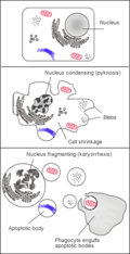 Apoptosis
 -
 4 Bd obs 4 680x512px
 -
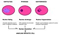 Nuclear changes
 -
 Histopathology of a pheochromocytoma with coagulative necrosis, with immunostaining
 -
 Apoptotic DNA Laddering


