Instruments used in radiology: Difference between revisions
CSV import |
CSV import |
||
| Line 31: | Line 31: | ||
[[Category:Medical imaging]] | [[Category:Medical imaging]] | ||
{{Medicine-stub}} | {{Medicine-stub}} | ||
<gallery> | |||
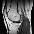
File:Knie mr.jpg|Instruments used in radiology | |||
File:Modern 3T MRI.JPG|Instruments used in radiology | |||
File:Varian4T.jpg|Instruments used in radiology | |||
File:Rontgenbuis-draaianode.jpg|Instruments used in radiology | |||
File:Laprascopy-Roentgen.jpg|Instruments used in radiology | |||
File:Mobile X-ray machine.jpg|Instruments used in radiology | |||
</gallery> | |||
Latest revision as of 01:35, 20 February 2025
Radiology is a medical specialty that uses imaging to diagnose and treat diseases within the body. A variety of imaging techniques such as X-ray radiography, Ultrasound, Computed Tomography (CT), Nuclear medicine including Positron Emission Tomography (PET) and Magnetic Resonance Imaging (MRI) are used to diagnose or treat diseases. Interventional radiology is the performance of medical procedures with the guidance of imaging technologies. The acquisition of medical images is usually carried out by the Radiographer, often known as a Radiologic Technologist. Depending on location, the Radiologist may interpret the images and write a report.
Instruments used in Radiology[edit]
X-ray Machine[edit]
An X-ray machine is one of the most common tools in radiology. It uses ionizing radiation to create images of the inside of the body. It is often used to view bones and can be used to diagnose fractures, infections, and tumors.
CT Scanner[edit]
A CT scanner uses X-rays and a computer to create detailed images of the inside of the body. It can provide more detailed information than standard X-rays and is often used to diagnose diseases or to plan medical treatments.
MRI Machine[edit]
An MRI machine uses a magnetic field and radio waves to create detailed images of the inside of the body. It is often used to diagnose conditions in the brain, spine, joints, and soft tissues.
Ultrasound Machine[edit]
An Ultrasound machine uses high-frequency sound waves to create images of the inside of the body. It is often used to view organs and structures within the body and is commonly used during pregnancy.
PET Scanner[edit]
A PET scanner uses a radioactive substance, called a tracer, to look for disease in the body. It is often used to detect cancer, heart problems, and brain disorders.
Nuclear Medicine Camera[edit]
A Nuclear medicine camera is used to create images of the body and certain diseases which are seen with a nuclear medicine study. It uses a small amount of radioactive material to diagnose, evaluate or treat a variety of diseases.
See Also[edit]

-
 Instruments used in radiology
 -
Instruments used in radiology
-
Instruments used in radiology
-
Instruments used in radiology
-
Instruments used in radiology
-
Instruments used in radiology

