Discoid meniscus: Difference between revisions
CSV import Tags: mobile edit mobile web edit |
CSV import |
||
| Line 28: | Line 28: | ||
[[Category:Knee injuries and disorders]] | [[Category:Knee injuries and disorders]] | ||
{{Medicine-stub}} | {{Medicine-stub}} | ||
== Discoid_meniscus == | |||
<gallery> | |||
File:Scheibenmeniscus.svg|Diagram of a discoid meniscus | |||
File:Normal_meniscus.png|Diagram of a normal meniscus | |||
File:CONGENITAL_DISCOID_MENISCI.jpg|Congenital discoid menisci | |||
File:Scheibenmeniskus_MRT_PDW_cor.jpg|MRI of a discoid meniscus | |||
</gallery> | |||
Revision as of 05:05, 18 February 2025
Discoid meniscus is a rare human anatomical variant that usually affects the knee joint. In this condition, the meniscus is thicker and disc-shaped, unlike the normal crescent-shaped meniscus. This anomaly is most commonly found in the lateral meniscus and can lead to a range of knee problems, including pain, swelling, and instability.
Anatomy
The meniscus is a fibrocartilaginous structure in the knee joint that acts as a shock absorber between the femur (thigh bone) and tibia (shin bone). There are two menisci in each knee: the medial meniscus and the lateral meniscus. The normal meniscus is crescent-shaped, but in a discoid meniscus, it is disc-shaped.
Epidemiology
Discoid meniscus is a rare condition, with a reported prevalence of 0.4% to 17% in the general population. It is more common in Asian populations and is often bilateral, meaning it affects both knees.
Clinical Presentation
Patients with discoid meniscus often present with knee pain, swelling, and instability. They may also experience a clicking or popping sensation in the knee. The symptoms usually begin in childhood or adolescence.
Diagnosis
The diagnosis of discoid meniscus is typically made using magnetic resonance imaging (MRI). This imaging technique can clearly show the shape and thickness of the meniscus, allowing for a definitive diagnosis.
Treatment
The treatment for discoid meniscus depends on the severity of the symptoms. Conservative treatment options include physical therapy and non-steroidal anti-inflammatory drugs (NSAIDs). If these treatments are not effective, surgical intervention may be necessary. The most common surgical procedure is a meniscectomy, in which the abnormal portion of the meniscus is removed.
Prognosis
The prognosis for discoid meniscus is generally good, especially if the condition is diagnosed and treated early. However, some patients may develop osteoarthritis in the affected knee later in life.

Discoid_meniscus
-
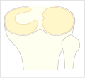 Diagram of a discoid meniscus
 -
Diagram of a normal meniscus
-
 Congenital discoid menisci
 -
 MRI of a discoid meniscus


