Falx cerebri: Difference between revisions
CSV import |
CSV import Tags: mobile edit mobile web edit |
||
| Line 27: | Line 27: | ||
{{medicine-stub}} | {{medicine-stub}} | ||
<gallery> | |||
File:Sobo_1909_589.png|Falx cerebri | |||
File:Gray135.png|Falx cerebri | |||
File:Gray568.png|Falx cerebri | |||
File:Human_brain_dura_mater_(reflections)_description.JPG|Falx cerebri | |||
File:Falx_cerebri.jpg|Falx cerebri | |||
</gallery> | |||
Latest revision as of 04:59, 18 February 2025
Falx Cerebri
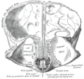
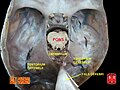
The falx cerebri is a dural fold that extends into the longitudinal fissure between the two cerebral hemispheres of the human brain. It is one of the dural septa, partitions of the dura mater, which is the outermost of the three layers of the meninges that surround the brain and spinal cord. The primary role of the falx cerebri is to help support and protect the brain by limiting its movement within the skull.
Structure[edit]
The falx cerebri is anchored anteriorly to the crista galli of the ethmoid bone and posteriorly to the internal occipital protuberance. It is narrow in front, where it is attached to the crista galli, and becomes wider as it progresses posteriorly. The inferior edge of the falx cerebri contains the superior sagittal sinus and the inferior sagittal sinus, which are important venous channels that drain blood from the brain back to the heart.
Function[edit]
The main function of the falx cerebri is to separate the two cerebral hemispheres and provide a supportive structure for the brain. By doing so, it plays a crucial role in protecting the brain from jarring movements that could cause injury. Additionally, the sinuses contained within the falx cerebri are essential for venous blood drainage from the brain.
Clinical Significance[edit]
- Intracranial Hemorrhage
The falx cerebri can be involved in certain types of intracranial hemorrhage, such as subdural hematoma, which occurs beneath the dura mater. Trauma to the head can lead to tearing of the veins that bridge the cerebral cortex and the dural sinuses, potentially causing blood to accumulate in this area.
- Falx Cerebri Tumors
Although rare, tumors can develop in the falx cerebri. These are usually meningiomas, which are typically benign tumors that arise from the meninges. Symptoms of a falx cerebri tumor can include headaches, seizures, and other neurological deficits, depending on the size and location of the tumor.
Diagnosis[edit]
Imaging techniques such as magnetic resonance imaging (MRI) and computed tomography (CT) scans are crucial for diagnosing conditions related to the falx cerebri. These imaging modalities can provide detailed images of the falx cerebri, helping to identify abnormalities such as tumors or signs of hemorrhage.
Treatment[edit]
Treatment for conditions affecting the falx cerebri depends on the underlying cause. For tumors, surgical removal is often necessary, especially if the tumor is causing symptoms. In the case of intracranial hemorrhage, treatment may involve managing the symptoms, monitoring the patient, and in some cases, surgical intervention to relieve pressure on the brain.
This medical article is a stub. You can help Wikipedia by adding missing information. |
-
 Falx cerebri
 -
 Falx cerebri
 -
 Falx cerebri
 -
 Falx cerebri
 -
 Falx cerebri


_description.JPG)

