Posterior superior alveolar artery: Difference between revisions
CSV import |
CSV import |
||
| Line 36: | Line 36: | ||
* [[Root canal treatment]] | * [[Root canal treatment]] | ||
{{dictionary-stub1}} | {{dictionary-stub1}} | ||
== Posterior_superior_alveolar_artery == | |||
<gallery> | |||
File:Gray511.png|Diagram of the maxillary artery and its branches | |||
File:Gray511.svg|Diagram of the maxillary artery and its branches (SVG version) | |||
File:Posterior_superior_alveolar_artery.png|Posterior superior alveolar artery | |||
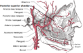
File:Gray157.png|Diagram of the arteries of the face and scalp | |||
</gallery> | |||
Latest revision as of 04:55, 18 February 2025
Posterior Superior Alveolar Artery[edit]
The posterior superior alveolar artery (PSAA) is a branch of the maxillary artery, which is one of the main arteries supplying blood to the upper jaw and its surrounding structures. The PSAA specifically provides blood supply to the maxillary molars and surrounding tissues in the posterior region of the maxilla.
Anatomy[edit]
The PSAA arises from the maxillary artery within the infratemporal fossa, which is a space located deep to the zygomatic arch. It typically originates just above the level of the maxillary tuberosity, which is the bony prominence at the back of the maxilla. From its origin, the PSAA courses superiorly and posteriorly towards the maxillary molars.
As it travels towards the posterior region of the maxilla, the PSAA gives off several branches. These branches include the dental branches, which supply blood to the dental pulp and periodontal tissues of the maxillary molars. Additionally, the PSAA may also give rise to branches that supply blood to the maxillary sinus, the buccal gingiva, and the surrounding bone.
Clinical Significance[edit]
The PSAA is of particular importance in dentistry, as it plays a crucial role in dental procedures involving the maxillary molars. Understanding the anatomy and variations of the PSAA is essential to minimize the risk of complications during these procedures.
During dental procedures such as tooth extraction or root canal treatment in the maxillary molars, the PSAA can be at risk of injury. Damage to the PSAA can result in bleeding and potential complications such as hematoma formation or compromised blood supply to the surrounding tissues. Therefore, it is important for dental professionals to be aware of the location and course of the PSAA to avoid inadvertent damage.
Variations[edit]
The anatomy of the PSAA can vary among individuals. One common variation is the presence of an accessory PSAA, which is an additional artery that accompanies the main PSAA. This accessory artery may arise from the maxillary artery or directly from the external carotid artery. The presence of an accessory PSAA can increase the risk of bleeding during dental procedures and should be taken into consideration when planning treatment.
References[edit]
1. Naitoh M, Hiraiwa Y, Aimiya H, et al. Arterial supply to the maxillary first molar examined by cone-beam computed tomography. Oral Surg Oral Med Oral Pathol Oral Radiol Endod. 2009;107(6):e34-e39. doi:10.1016/j.tripleo.2009.01.034
2. Kawai T, Asaumi R, Sato I, et al. Assessment of the posterior superior alveolar artery and the maxillary sinus with cone-beam computed tomography. Oral Surg Oral Med Oral Pathol Oral Radiol Endod. 2009;107(2):e26-e32. doi:10.1016/j.tripleo.2008.10.014
3. Kim S, Kim E, Song H, et al. Anatomical characteristics and variations of the posterior superior alveolar artery. Clin Oral Implants Res. 2013;24(4):416-421. doi:10.1111/j.1600-0501.2011.02384.x
See Also[edit]

Posterior_superior_alveolar_artery[edit]
-
 Diagram of the maxillary artery and its branches
 -
 Diagram of the maxillary artery and its branches (SVG version)
 -
 Posterior superior alveolar artery
 -
 Diagram of the arteries of the face and scalp


