Focus assessed transthoracic echocardiography: Difference between revisions
CSV import |
CSV import |
||
| Line 23: | Line 23: | ||
{{medicine-stub}} | {{medicine-stub}} | ||
<gallery> | |||
File:Subcostal_view_of_heart.gif|Subcostal view of heart | |||
File:Apical_4_chamber_view.png|Apical 4 chamber view | |||
File:LeftParasternalLongAxis.gif|Left parasternal long axis | |||
File:LeftVentricleShortAxis.gif|Left ventricle short axis | |||
</gallery> | |||
Latest revision as of 04:45, 18 February 2025
Focus Assessed Transthoracic Echocardiography (FATE) is a medical imaging technique used to visualize the heart and its surrounding structures. It is a type of echocardiography that is specifically designed to provide a focused assessment of the heart's structure and function.
Overview[edit]
FATE is a non-invasive procedure that uses ultrasound waves to create images of the heart. It is commonly used in the emergency department, intensive care unit, and during surgery to quickly assess the heart's function and to guide treatment decisions.
Procedure[edit]
During a FATE examination, the patient lies on their back and the sonographer applies a gel to the chest. The sonographer then places an ultrasound probe on the chest and moves it around to obtain images of the heart from different angles. The images are displayed on a monitor for the sonographer and physician to review.
Indications[edit]
FATE is used to assess the heart's structure and function in patients with a variety of conditions, including heart failure, myocardial infarction, and cardiac tamponade. It can also be used to guide procedures such as pericardiocentesis and to monitor the heart during surgery.
Limitations[edit]
While FATE is a valuable tool in the assessment of the heart, it does have some limitations. It is dependent on the skill of the sonographer, and the quality of the images can be affected by factors such as the patient's body habitus and lung disease. Additionally, FATE is a focused examination and may not provide a comprehensive assessment of the heart.
See Also[edit]

-
 Subcostal view of heart
 -
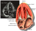 Apical 4 chamber view
 -
 Left parasternal long axis
 -
 Left ventricle short axis


