Magnetic resonance elastography: Difference between revisions
CSV import |
CSV import |
||
| Line 30: | Line 30: | ||
[[Category:Magnetic resonance imaging]] | [[Category:Magnetic resonance imaging]] | ||
{{medicine-stub}} | {{medicine-stub}} | ||
<gallery> | |||
File:LiverMRE_wiki_final.tif|Magnetic resonance elastography of the liver | |||
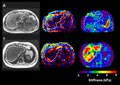
File:Murphy_2013_brain_MRE_with_wave_image.png|Brain magnetic resonance elastography with wave image | |||
File:MRE-of_Kidney_Prostate_and_Pancreas.tif|Magnetic resonance elastography of kidney, prostate, and pancreas | |||
</gallery> | |||
Latest revision as of 04:08, 18 February 2025
Magnetic Resonance Elastography (MRE) is a non-invasive imaging technique that measures the stiffness of soft tissues in the body. It is an advanced application of Magnetic Resonance Imaging (MRI) technology, combining traditional MRI images with information about the mechanical properties of tissues. This technique is particularly useful in the diagnosis and assessment of diseases in organs such as the liver, brain, and breast, where tissue stiffness can indicate the presence of disease.
Overview[edit]
Magnetic Resonance Elastography works by generating mechanical waves within the tissue using an external driver. These waves are then imaged by the MRI machine as they propagate through the tissue. The data collected is processed to generate a quantitative map of tissue stiffness, known as an elastogram. The stiffness of the tissue can indicate the presence of fibrosis, tumors, or other pathologies.
Applications[edit]
MRE is most commonly used in the diagnosis and staging of liver fibrosis and cirrhosis. It has proven to be a reliable and accurate method for assessing liver stiffness, which correlates with the degree of fibrosis. This is particularly important for the management of chronic liver diseases, such as hepatitis C and non-alcoholic fatty liver disease (NAFLD), where the degree of fibrosis is a critical factor in treatment decisions.
In addition to liver applications, MRE is also being explored for use in other organs:
- In the brain, MRE can help in the diagnosis of diseases such as Alzheimer's disease, multiple sclerosis, and brain tumors.
- In the breast, it has potential for improving the specificity of MRI in the differentiation of benign and malignant lesions.
- MRE is also being investigated for its utility in the heart, kidneys, and prostate.
Advantages[edit]
MRE has several advantages over traditional imaging and biopsy methods:
- It is non-invasive, avoiding the risks associated with tissue biopsies.
- It provides a comprehensive view of the entire organ, as opposed to a biopsy which only samples a small area.
- It can quantitatively measure tissue stiffness, providing objective data that can be tracked over time.
Limitations[edit]
While MRE is a powerful tool, it also has limitations:
- It requires specialized equipment and expertise, which may not be available in all medical facilities.
- It can be more expensive than other imaging modalities.
- Patients with certain types of implants or devices may not be suitable for MRI exams, including MRE.
Future Directions[edit]
Research is ongoing to expand the applications of MRE and to refine the technology. This includes developing faster and more sensitive imaging techniques, as well as exploring the use of MRE in a wider range of tissues and diseases.

-
 Magnetic resonance elastography of the liver
 -
 Brain magnetic resonance elastography with wave image
 -
 Magnetic resonance elastography of kidney, prostate, and pancreas

