Trochanter: Difference between revisions
CSV import Tags: mobile edit mobile web edit |
CSV import |
||
| Line 1: | Line 1: | ||
= | {{Short description|A part of the human femur bone}} | ||
{{Use dmy dates|date=October 2023}} | |||
The trochanter is a bony prominence | ==Trochanter== | ||
The '''trochanter''' is a bony prominence near the proximal end of the femur, the bone of the thigh. In humans, there are two trochanters: the greater trochanter and the lesser trochanter. These structures serve as important sites for muscle attachment and play a crucial role in the movement and stability of the hip joint. | |||
=== | ===Greater Trochanter=== | ||
The '''greater trochanter''' is a large, irregular, quadrilateral eminence located at the junction of the neck and shaft of the femur. It is palpable under the skin and serves as an attachment point for several muscles, including the [[gluteus medius]], [[gluteus minimus]], and [[piriformis]]. The greater trochanter is a key landmark in orthopedic surgery and is often used as a reference point in hip replacement procedures. | |||
The femur | ===Lesser Trochanter=== | ||
The '''lesser trochanter''' is a smaller, conical projection located on the medial side of the femur, below the neck. It serves as the insertion point for the [[iliopsoas]] muscle, which is a major flexor of the hip joint. The lesser trochanter is less prominent than the greater trochanter but is equally important for muscle attachment and function. | |||
The | ==Function== | ||
The trochanters are essential for the attachment of muscles that move the hip joint. The greater trochanter provides leverage for the muscles that abduct and rotate the thigh, while the lesser trochanter is involved in flexing the hip. These bony prominences also help stabilize the hip joint during movement, contributing to the overall biomechanics of walking and running. | |||
==Clinical Significance== | |||
Injuries or conditions affecting the trochanters can lead to significant pain and mobility issues. Trochanteric bursitis, for example, is a common condition characterized by inflammation of the bursa near the greater trochanter, leading to hip pain. Fractures involving the trochanters, such as intertrochanteric fractures, are also common, especially in the elderly population, and require surgical intervention. | |||
==Related Pages== | |||
* [[Femur]] | * [[Femur]] | ||
* [[Hip | * [[Hip joint]] | ||
* [[ | * [[Musculoskeletal system]] | ||
== | ==Gallery== | ||
<gallery> | |||
File:Gray243.png|Diagram of the femur showing the greater and lesser trochanters. | |||
File:Replica_of_Athenian_trireme_(trieres)._Athens_War_Museum.jpg|A replica of an Athenian trireme, illustrating the historical context of the term "trochanter" in ancient Greek. | |||
</gallery> | |||
==References== | |||
* Gray, Henry. ''Anatomy of the Human Body''. 20th ed. Philadelphia: Lea & Febiger, 1918. | |||
* Standring, Susan, ed. ''Gray's Anatomy: The Anatomical Basis of Clinical Practice''. 41st ed. Elsevier, 2016. | |||
[[Category:Anatomy | [[Category:Anatomy of the lower limb]] | ||
Revision as of 20:59, 9 February 2025
A part of the human femur bone
Trochanter
The trochanter is a bony prominence near the proximal end of the femur, the bone of the thigh. In humans, there are two trochanters: the greater trochanter and the lesser trochanter. These structures serve as important sites for muscle attachment and play a crucial role in the movement and stability of the hip joint.
Greater Trochanter
The greater trochanter is a large, irregular, quadrilateral eminence located at the junction of the neck and shaft of the femur. It is palpable under the skin and serves as an attachment point for several muscles, including the gluteus medius, gluteus minimus, and piriformis. The greater trochanter is a key landmark in orthopedic surgery and is often used as a reference point in hip replacement procedures.
Lesser Trochanter
The lesser trochanter is a smaller, conical projection located on the medial side of the femur, below the neck. It serves as the insertion point for the iliopsoas muscle, which is a major flexor of the hip joint. The lesser trochanter is less prominent than the greater trochanter but is equally important for muscle attachment and function.
Function
The trochanters are essential for the attachment of muscles that move the hip joint. The greater trochanter provides leverage for the muscles that abduct and rotate the thigh, while the lesser trochanter is involved in flexing the hip. These bony prominences also help stabilize the hip joint during movement, contributing to the overall biomechanics of walking and running.
Clinical Significance
Injuries or conditions affecting the trochanters can lead to significant pain and mobility issues. Trochanteric bursitis, for example, is a common condition characterized by inflammation of the bursa near the greater trochanter, leading to hip pain. Fractures involving the trochanters, such as intertrochanteric fractures, are also common, especially in the elderly population, and require surgical intervention.
Related Pages
Gallery
-
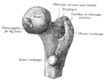 Diagram of the femur showing the greater and lesser trochanters.
 -
 A replica of an Athenian trireme, illustrating the historical context of the term "trochanter" in ancient Greek.

._Athens_War_Museum.jpg)
References
- Gray, Henry. Anatomy of the Human Body. 20th ed. Philadelphia: Lea & Febiger, 1918.
- Standring, Susan, ed. Gray's Anatomy: The Anatomical Basis of Clinical Practice. 41st ed. Elsevier, 2016.
