Articulation of head of rib: Difference between revisions
CSV import |
CSV import |
||
| Line 37: | Line 37: | ||
[[Category:Neurosurgery]] | [[Category:Neurosurgery]] | ||
[[Category:Congenital disorders]] | [[Category:Congenital disorders]] | ||
<gallery> | |||
File:Gray312.png|Articulation of head of rib | |||
File:Gray90.png|Articulation of head of rib | |||
File:Gray204.png|Articulation of head of rib | |||
</gallery> | |||
Revision as of 01:18, 18 February 2025
Aqueductal Stenosis
Aqueductal stenosis is a condition characterized by the narrowing of the cerebral aqueduct, which is a channel that connects the third ventricle to the fourth ventricle in the brain. This narrowing can lead to an obstruction of the flow of cerebrospinal fluid (CSF), resulting in a condition known as hydrocephalus.
Anatomy and Physiology
The cerebral aqueduct, also known as the aqueduct of Sylvius, is a slender canal located within the midbrain. It is part of the ventricular system of the brain, which is responsible for the production, transport, and removal of cerebrospinal fluid. The aqueduct is approximately 1.5 mm in diameter and is crucial for the passage of CSF from the third to the fourth ventricle.
Pathophysiology
Aqueductal stenosis can be congenital or acquired. Congenital aqueductal stenosis is often due to developmental anomalies, such as aqueductal webs or atresia. Acquired stenosis may result from infections, hemorrhage, or tumors that compress or invade the aqueduct.
The obstruction of CSF flow leads to increased pressure within the ventricular system, causing the ventricles to enlarge. This condition, known as hydrocephalus, can result in increased intracranial pressure, which may cause symptoms such as headaches, nausea, vomiting, and in severe cases, brain herniation.
Clinical Presentation
Patients with aqueductal stenosis may present with a variety of symptoms depending on the age of onset and the severity of the obstruction. In infants, it may cause an increase in head size, irritability, and poor feeding. In older children and adults, symptoms may include headaches, nausea, vomiting, balance problems, and cognitive difficulties.
Diagnosis
Diagnosis of aqueductal stenosis is typically made using neuroimaging techniques such as MRI or CT scan. These imaging modalities can reveal the enlargement of the ventricles and the narrowing of the aqueduct.
Treatment
The primary treatment for aqueductal stenosis is surgical intervention to relieve the obstruction and restore normal CSF flow. This can be achieved through procedures such as endoscopic third ventriculostomy (ETV) or the placement of a ventriculoperitoneal shunt. ETV involves creating an opening in the floor of the third ventricle to allow CSF to bypass the obstruction, while a shunt diverts excess fluid to another part of the body where it can be absorbed.
Related Pages
Gallery
-
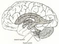 Diagram of the ventricular system showing the cerebral aqueduct.
 -
 Image of a hydrocephalic brain.
 -
 Animation of the third ventricle.
 -
 Animation of the fourth ventricle.


-
 Articulation of head of rib
 -
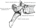 Articulation of head of rib
 -
 Articulation of head of rib


