Large-cell acanthoma: Difference between revisions
CSV import Tags: mobile edit mobile web edit |
CSV import |
||
| Line 31: | Line 31: | ||
{{Medicine-stub}} | {{Medicine-stub}} | ||
{{No image}} | {{No image}} | ||
<gallery> | |||
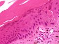
File:SkinTumors-P6120248.JPG|Large-cell acanthoma | |||
</gallery> | |||
Revision as of 01:40, 20 February 2025
Large-cell acanthoma is a benign skin lesion that is characterized by its large, pale-staining keratinocytes. It is a rare condition that is often mistaken for other skin conditions, such as actinic keratosis or squamous cell carcinoma.
History
Large-cell acanthoma was first described in 1968 by the dermatologist Robert Degos. It was initially thought to be a variant of seborrheic keratosis, but further studies have shown that it is a distinct entity.
Characteristics
Large-cell acanthoma typically presents as a solitary, well-demarcated, flat or slightly raised lesion. It is usually found on sun-exposed areas of the body, such as the face, neck, and arms. The lesion is often asymptomatic, but it may cause mild itching or discomfort.
Histologically, large-cell acanthoma is characterized by an increased number of large, pale-staining keratinocytes in the epidermis. These cells are larger than the surrounding keratinocytes and have a clear or pale cytoplasm. The dermis underneath the lesion may show signs of solar elastosis, indicating chronic sun damage.
Diagnosis
The diagnosis of large-cell acanthoma is usually made based on the clinical appearance of the lesion and the histological findings. A skin biopsy may be performed to confirm the diagnosis and to rule out other skin conditions.
Treatment
Treatment for large-cell acanthoma is usually not necessary, as the condition is benign and does not pose a risk for malignancy. However, if the lesion is bothersome or cosmetically unacceptable, it can be removed by surgical excision or cryotherapy.
See also
This dermatology article is a stub. You can help WikiMD by expanding the page. |
This medical article is a stub. You can help WikiMD by expanding the page. |
-
 Large-cell acanthoma

