Parietal bone: Difference between revisions
CSV import |
CSV import |
||
| (One intermediate revision by the same user not shown) | |||
| Line 1: | Line 1: | ||
{{Short description|Bone forming part of the side and top of the skull}} | |||
{{Use dmy dates|date=October 2023}} | |||
The '''parietal bone''' is a bone | ==Parietal bone== | ||
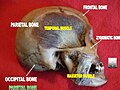
The '''parietal bone''' is a paired, flat bone located on each side of the [[human skull]]. It forms a large part of the [[calvaria]], or skullcap, and is situated between the [[frontal bone]] and the [[occipital bone]]. | |||
== | [[File:Parietal_bone_posterior2.png|thumb|right|Posterior view of the parietal bone]] | ||
The parietal bone articulates with the [[frontal bone]] | |||
==Anatomy== | |||
The parietal bone is quadrilateral in shape and has four borders, four angles, and two surfaces. It articulates with five other bones: the [[frontal bone]], [[occipital bone]], [[temporal bone]], and the opposite parietal bone. | |||
===Borders=== | ===Borders=== | ||
The parietal bone | * '''Sagittal border''': The superior border, which articulates with the opposite parietal bone at the [[sagittal suture]]. | ||
* '''Squamous border''': The inferior border, which articulates with the [[temporal bone]]. | |||
* '''Frontal border''': The anterior border, which articulates with the [[frontal bone]] at the [[coronal suture]]. | |||
* '''Occipital border''': The posterior border, which articulates with the [[occipital bone]] at the [[lambdoid suture]]. | |||
[[File:Sagittal_suture_2.jpg|thumb|left|The sagittal suture between the two parietal bones]] | |||
===Angles=== | ===Angles=== | ||
* '''Frontal angle''': Located at the junction of the coronal and sagittal sutures. | |||
* '''Sphenoidal angle''': Located at the junction of the coronal and squamous sutures. | |||
* '''Occipital angle''': Located at the junction of the sagittal and lambdoid sutures. | |||
* '''Mastoid angle''': Located at the junction of the squamous and lambdoid sutures. | |||
* | ===Surfaces=== | ||
* | * '''External surface''': Convex and smooth, providing attachment for the [[temporalis muscle]]. | ||
* '''Internal surface''': Concave, with grooves for the [[middle meningeal artery]] and [[sagittal sinus]]. | |||
[[File:Gray132.png|thumb|right|Outer surface of the parietal bone]] | |||
[[File:Gray133.png|thumb|left|Inner surface of the parietal bone]] | |||
==Development== | ==Development== | ||
The parietal bone | The parietal bone develops from two primary ossification centers that appear in the membrane covering the developing brain. These centers appear around the eighth week of fetal development and fuse to form a single bone by birth. | ||
==Function== | |||
The parietal bone plays a crucial role in protecting the brain and forming the shape of the head. It also provides attachment points for muscles involved in mastication and head movement. | |||
==Clinical significance== | ==Clinical significance== | ||
Fractures of the parietal bone can occur due to trauma and may lead to complications such as [[epidural hematoma]]. The bone's thinness makes it susceptible to injury, but its position provides some protection. | |||
[[File:Left_parietal_boen_-_animation.gif|thumb|right|Animation showing the left parietal bone]] | |||
==Comparative anatomy== | |||
In other vertebrates, the parietal bone can vary significantly in size and shape. In some species, it may be fused with other bones or have additional functions. | |||
[[File:Tuatara_skull_diagram.svg|thumb|left|Diagram of a tuatara skull showing the parietal bone]] | |||
== | ==Gallery== | ||
< | <gallery> | ||
File:Parietal_bone_animation2.gif|Animation of the parietal bone | |||
File:Parietal_bone.jpg|Lateral view of the parietal bone | |||
File:HSCA-JFK-head-7-125.jpg|X-ray showing the parietal bone | |||
</gallery> | |||
== | ==Related pages== | ||
* [ | * [[Frontal bone]] | ||
* [[Occipital bone]] | |||
* [[Temporal bone]] | |||
* [[Sphenoid bone]] | |||
[[Category:Skull]] | [[Category:Skull]] | ||
[[Category:Bones of the head and neck]] | [[Category:Bones of the head and neck]] | ||
Latest revision as of 14:13, 21 February 2025
Bone forming part of the side and top of the skull
Parietal bone[edit]
The parietal bone is a paired, flat bone located on each side of the human skull. It forms a large part of the calvaria, or skullcap, and is situated between the frontal bone and the occipital bone.

Anatomy[edit]
The parietal bone is quadrilateral in shape and has four borders, four angles, and two surfaces. It articulates with five other bones: the frontal bone, occipital bone, temporal bone, and the opposite parietal bone.
Borders[edit]
- Sagittal border: The superior border, which articulates with the opposite parietal bone at the sagittal suture.
- Squamous border: The inferior border, which articulates with the temporal bone.
- Frontal border: The anterior border, which articulates with the frontal bone at the coronal suture.
- Occipital border: The posterior border, which articulates with the occipital bone at the lambdoid suture.

Angles[edit]
- Frontal angle: Located at the junction of the coronal and sagittal sutures.
- Sphenoidal angle: Located at the junction of the coronal and squamous sutures.
- Occipital angle: Located at the junction of the sagittal and lambdoid sutures.
- Mastoid angle: Located at the junction of the squamous and lambdoid sutures.
Surfaces[edit]
- External surface: Convex and smooth, providing attachment for the temporalis muscle.
- Internal surface: Concave, with grooves for the middle meningeal artery and sagittal sinus.


Development[edit]
The parietal bone develops from two primary ossification centers that appear in the membrane covering the developing brain. These centers appear around the eighth week of fetal development and fuse to form a single bone by birth.
Function[edit]
The parietal bone plays a crucial role in protecting the brain and forming the shape of the head. It also provides attachment points for muscles involved in mastication and head movement.
Clinical significance[edit]
Fractures of the parietal bone can occur due to trauma and may lead to complications such as epidural hematoma. The bone's thinness makes it susceptible to injury, but its position provides some protection.

Comparative anatomy[edit]
In other vertebrates, the parietal bone can vary significantly in size and shape. In some species, it may be fused with other bones or have additional functions.

Gallery[edit]
-
 Animation of the parietal bone
 -
 Lateral view of the parietal bone
 -
 X-ray showing the parietal bone


