Two-photon excitation microscopy: Difference between revisions
CSV import |
CSV import |
||
| Line 33: | Line 33: | ||
[[Category:Fluorescence techniques]] | [[Category:Fluorescence techniques]] | ||
[[Category:Biological imaging]] | [[Category:Biological imaging]] | ||
== Two-photon_excitation_microscopy == | |||
<gallery> | |||
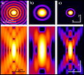
File:MultiPhotonExcitation-Fig10-doi10.1186slash1475-925X-5-36-clipping.JPEG|Two-photon_excitation_microscopy | |||
File:MultiPhotonExcitation-Fig1-doi10.1186slash1475-925X-5-36.JPEG|Two-photon_excitation_microscopy | |||
File:MultiPhotonExcitation-Fig7-doi10.1186slash1475-925X-5-36.JPEG|Two-photon_excitation_microscopy | |||
File:Diagram_of_a_two-photon_excitation_microscope_en.svg|Diagram of a two-photon excitation microscope | |||
File:Two-photon_microscopy_of_in_vivo_brain_function.jpg|Two-photon microscopy of in vivo brain function | |||
</gallery> | |||
Latest revision as of 11:27, 18 February 2025
Two-photon excitation microscopy[edit]


Two-photon excitation microscopy is a fluorescence imaging technique that allows imaging of living tissue up to a very high depth. It is a special variant of fluorescence microscopy that uses two photons of lower energy to excite a fluorophore, instead of one photon of higher energy. This technique was first developed in the 1990s and has since become a powerful tool in biological research.
Principle[edit]
Two-photon excitation relies on the simultaneous absorption of two photons by a fluorophore. Each photon has approximately half the energy (and thus twice the wavelength) required for excitation. This process is nonlinear and occurs only at the focal point of the laser, which allows for precise spatial localization of the excitation. The use of longer wavelengths reduces scattering in biological tissues, enabling deeper penetration and less photodamage compared to single-photon excitation.
Applications[edit]
Two-photon microscopy is widely used in neuroscience, cell biology, and developmental biology. It is particularly useful for imaging thick specimens, such as brain slices or whole embryos, where traditional fluorescence microscopy would be limited by scattering and absorption. The technique allows researchers to observe dynamic processes in living tissues, such as neuronal activity, blood flow, and cellular interactions.
Advantages[edit]
The main advantages of two-photon excitation microscopy include reduced photobleaching and phototoxicity, as well as improved imaging depth. The localized excitation reduces out-of-focus fluorescence, enhancing image contrast and resolution. Additionally, the use of infrared light minimizes damage to living tissues, making it ideal for long-term imaging studies.
Limitations[edit]
Despite its advantages, two-photon microscopy has some limitations. The requirement for high-intensity pulsed lasers can be costly and complex to operate. The technique also has lower temporal resolution compared to some other imaging methods, which can be a limitation for certain dynamic studies.
Related pages[edit]
References[edit]
<references group="" responsive="1"></references>
Two-photon_excitation_microscopy[edit]
-
 Two-photon_excitation_microscopy
 -
 Two-photon_excitation_microscopy
 -
 Two-photon_excitation_microscopy
 -
 Diagram of a two-photon excitation microscope
 -
 Two-photon microscopy of in vivo brain function
