Trochlear nerve: Difference between revisions
CSV import Tags: mobile edit mobile web edit |
No edit summary |
||
| (2 intermediate revisions by the same user not shown) | |||
| Line 1: | Line 1: | ||
''' | {{Short description|Fourth cranial nerve of the human body}} | ||
The '''trochlear nerve''' is the fourth cranial nerve (CN IV) and is a motor nerve that innervates the superior oblique muscle of the eye, which controls rotational movement. It is unique among the cranial nerves in that it is the only one that emerges dorsally from the brainstem and the only one that decussates (crosses to the opposite side) before innervating its target muscle. | |||
== Anatomy == | ==Anatomy== | ||
The trochlear nerve originates in the midbrain, specifically from the trochlear nucleus, which is located at the level of the inferior colliculus. It emerges from the dorsal aspect of the brainstem, just below the inferior colliculus, and then wraps around the brainstem to reach the ventral side. | |||
[[File:Brain_human_normal_inferior_view_with_labels_en.svg|thumb|Inferior view of the human brain, showing the location of the trochlear nerve.]] | |||
After emerging from the brainstem, the trochlear nerve travels anteriorly within the subarachnoid space, passing between the posterior cerebral artery and the superior cerebellar artery. It then pierces the dura mater to enter the cavernous sinus, where it runs along the lateral wall. Finally, it enters the orbit through the superior orbital fissure to innervate the superior oblique muscle. | |||
The | ==Function== | ||
The primary function of the trochlear nerve is to provide motor innervation to the superior oblique muscle. This muscle is responsible for intorsion (inward rotation), depression, and abduction of the eye. The unique action of the superior oblique muscle allows for the downward and outward movement of the eye, which is essential for proper binocular vision and depth perception. | |||
== Clinical significance == | ==Clinical significance== | ||
Damage to the trochlear nerve can result in a condition known as [[trochlear nerve palsy]]. This condition is characterized by vertical diplopia (double vision) and difficulty in moving the eye downward, especially when looking towards the nose. Patients may compensate for this by tilting their head to the opposite side of the affected eye. | |||
[[File:Gray571.png|thumb|Diagram of the cranial nerve nuclei, showing the trochlear nerve nucleus.]] | |||
Causes of trochlear nerve palsy can include trauma, microvascular disease, congenital defects, or tumors. Diagnosis is typically made through clinical examination and imaging studies, such as MRI or CT scans. | |||
== Gallery == | |||
<gallery> | |||
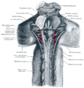
File:Trochlear_nerve.png|Trochlear nerve | |||
File:Gray571.png|Diagram of the cranial nerves | |||
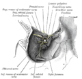
File:Gray719.png|Nerves of the orbit | |||
File:Gray787.png|The cranial nerve nuclei | |||
File:Gray792.png|The position of the trochlear nerve | |||
File:Slide2ior.JPG|Trochlear nerve | |||
</gallery> | |||
==Related pages== | |||
* [[Cranial nerves]] | * [[Cranial nerves]] | ||
* [[Superior oblique muscle]] | * [[Superior oblique muscle]] | ||
* [[ | * [[Diplopia]] | ||
[[Category:Cranial nerves]] | [[Category:Cranial nerves]] | ||
Latest revision as of 03:22, 28 March 2025
Fourth cranial nerve of the human body
The trochlear nerve is the fourth cranial nerve (CN IV) and is a motor nerve that innervates the superior oblique muscle of the eye, which controls rotational movement. It is unique among the cranial nerves in that it is the only one that emerges dorsally from the brainstem and the only one that decussates (crosses to the opposite side) before innervating its target muscle.
Anatomy[edit]
The trochlear nerve originates in the midbrain, specifically from the trochlear nucleus, which is located at the level of the inferior colliculus. It emerges from the dorsal aspect of the brainstem, just below the inferior colliculus, and then wraps around the brainstem to reach the ventral side.

After emerging from the brainstem, the trochlear nerve travels anteriorly within the subarachnoid space, passing between the posterior cerebral artery and the superior cerebellar artery. It then pierces the dura mater to enter the cavernous sinus, where it runs along the lateral wall. Finally, it enters the orbit through the superior orbital fissure to innervate the superior oblique muscle.
Function[edit]
The primary function of the trochlear nerve is to provide motor innervation to the superior oblique muscle. This muscle is responsible for intorsion (inward rotation), depression, and abduction of the eye. The unique action of the superior oblique muscle allows for the downward and outward movement of the eye, which is essential for proper binocular vision and depth perception.
Clinical significance[edit]
Damage to the trochlear nerve can result in a condition known as trochlear nerve palsy. This condition is characterized by vertical diplopia (double vision) and difficulty in moving the eye downward, especially when looking towards the nose. Patients may compensate for this by tilting their head to the opposite side of the affected eye.
Causes of trochlear nerve palsy can include trauma, microvascular disease, congenital defects, or tumors. Diagnosis is typically made through clinical examination and imaging studies, such as MRI or CT scans.
Gallery[edit]
-
 Trochlear nerve
 -
 Diagram of the cranial nerves
 -
 Nerves of the orbit
 -
 The cranial nerve nuclei
 -
 The position of the trochlear nerve
 -
 Trochlear nerve


