Tympanic cavity: Difference between revisions
CSV import |
Tag: Manual revert |
||
| (2 intermediate revisions by the same user not shown) | |||
| Line 1: | Line 1: | ||
{{Short description|Anatomical cavity in the ear}} | |||
{{Use dmy dates|date=October 2023}} | |||
The ''' | The '''tympanic cavity''' is a small, air-filled space located in the [[temporal bone]] of the [[skull]]. It is part of the [[middle ear]] and plays a crucial role in the process of hearing by transmitting sound vibrations from the [[eardrum]] to the [[inner ear]]. | ||
==Anatomy== | ==Anatomy== | ||
The tympanic cavity is bounded laterally by the [[tympanic membrane]] (eardrum) and medially by the [[bony labyrinth]] of the inner ear. It is connected to the [[nasopharynx]] via the [[Eustachian tube]], which helps equalize pressure between the middle ear and the atmosphere. | |||
The tympanic cavity | ===Walls=== | ||
The tympanic cavity has six walls: | |||
* '''Lateral wall''': Formed by the tympanic membrane. | |||
* '''Medial wall''': Contains the [[oval window]] and [[round window]], which are openings into the inner ear. | |||
* '''Anterior wall''': Contains the opening of the Eustachian tube and the canal for the [[tensor tympani muscle]]. | |||
* '''Posterior wall''': Contains the entrance to the [[mastoid antrum]] and the [[pyramidal eminence]]. | |||
* '''Roof''': Formed by a thin plate of bone called the [[tegmen tympani]]. | |||
* '''Floor''': Formed by the jugular wall, which separates the tympanic cavity from the [[jugular fossa]]. | |||
=== | ===Contents=== | ||
The tympanic cavity contains three small bones known as the [[ossicles]]: the [[malleus]], [[incus]], and [[stapes]]. These bones form a chain that transmits sound vibrations from the tympanic membrane to the oval window of the inner ear. | |||
The tympanic cavity | |||
==Function== | ==Function== | ||
The primary function of the tympanic cavity is to facilitate the transmission of sound from the external ear to the inner ear. The ossicles amplify and convey sound vibrations from the tympanic membrane to the oval window, where they are converted into fluid waves in the cochlea of the inner ear. | |||
==Clinical significance== | |||
Conditions affecting the tympanic cavity include [[otitis media]], which is an infection or inflammation of the middle ear, and [[otosclerosis]], a condition that affects the movement of the stapes bone. Proper functioning of the Eustachian tube is essential for maintaining equal air pressure on both sides of the tympanic membrane. | |||
== | ==Images== | ||
<gallery> | |||
File:Gray923.png|Diagram of the tympanic cavity. | |||
File:Gray907.png|Ossicles of the middle ear. | |||
File:Gray908.png|Tympanic cavity and surrounding structures. | |||
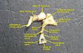
File:Occipital_bone_dissection.jpg|Dissection showing the temporal bone. | |||
File:Slide1ghe.JPG|View of the tympanic cavity. | |||
</gallery> | |||
==Related pages== | |||
* [[Middle ear]] | |||
* [[Eustachian tube]] | |||
* [[Ossicles]] | |||
* [[Tympanic membrane]] | |||
== | ==References== | ||
* Moore, K. L., Dalley, A. F., & Agur, A. M. R. (2013). ''Clinically Oriented Anatomy''. Lippincott Williams & Wilkins. | |||
* Standring, S. (2015). ''Gray's Anatomy: The Anatomical Basis of Clinical Practice''. Elsevier Health Sciences. | |||
[[Category:Auditory system]] | [[Category:Auditory system]] | ||
[[Category:Human head and neck]] | |||
Latest revision as of 13:41, 22 March 2025
Anatomical cavity in the ear
The tympanic cavity is a small, air-filled space located in the temporal bone of the skull. It is part of the middle ear and plays a crucial role in the process of hearing by transmitting sound vibrations from the eardrum to the inner ear.
Anatomy[edit]
The tympanic cavity is bounded laterally by the tympanic membrane (eardrum) and medially by the bony labyrinth of the inner ear. It is connected to the nasopharynx via the Eustachian tube, which helps equalize pressure between the middle ear and the atmosphere.
Walls[edit]
The tympanic cavity has six walls:
- Lateral wall: Formed by the tympanic membrane.
- Medial wall: Contains the oval window and round window, which are openings into the inner ear.
- Anterior wall: Contains the opening of the Eustachian tube and the canal for the tensor tympani muscle.
- Posterior wall: Contains the entrance to the mastoid antrum and the pyramidal eminence.
- Roof: Formed by a thin plate of bone called the tegmen tympani.
- Floor: Formed by the jugular wall, which separates the tympanic cavity from the jugular fossa.
Contents[edit]
The tympanic cavity contains three small bones known as the ossicles: the malleus, incus, and stapes. These bones form a chain that transmits sound vibrations from the tympanic membrane to the oval window of the inner ear.
Function[edit]
The primary function of the tympanic cavity is to facilitate the transmission of sound from the external ear to the inner ear. The ossicles amplify and convey sound vibrations from the tympanic membrane to the oval window, where they are converted into fluid waves in the cochlea of the inner ear.
Clinical significance[edit]
Conditions affecting the tympanic cavity include otitis media, which is an infection or inflammation of the middle ear, and otosclerosis, a condition that affects the movement of the stapes bone. Proper functioning of the Eustachian tube is essential for maintaining equal air pressure on both sides of the tympanic membrane.
Images[edit]
-
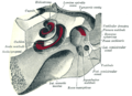 Diagram of the tympanic cavity.
 -
 Ossicles of the middle ear.
 -
 Tympanic cavity and surrounding structures.
 -
 Dissection showing the temporal bone.
 -
 View of the tympanic cavity.


Related pages[edit]
References[edit]
- Moore, K. L., Dalley, A. F., & Agur, A. M. R. (2013). Clinically Oriented Anatomy. Lippincott Williams & Wilkins.
- Standring, S. (2015). Gray's Anatomy: The Anatomical Basis of Clinical Practice. Elsevier Health Sciences.
