Cochlear duct: Difference between revisions
CSV import |
CSV import |
||
| Line 29: | Line 29: | ||
[[Category:Hearing]] | [[Category:Hearing]] | ||
{{anatomy-stub}} | {{anatomy-stub}} | ||
<gallery> | |||
File:Blausen_0329_EarAnatomy_InternalEar.png|Diagram of the internal ear anatomy | |||
File:Gray928.png|Cross-section of the cochlear duct | |||
File:Gray903.png|Anatomy of the cochlea | |||
File:Gray924.png|Structure of the cochlear duct | |||
File:Gray929.png|Detailed view of the cochlear duct | |||
File:Cochlea-crosssection.svg|Cross-section of the cochlea | |||
</gallery> | |||
Latest revision as of 05:02, 18 February 2025
Cochlear Duct
The cochlear duct or scala media is a component of the inner ear that plays a crucial role in the process of hearing. It is a part of the cochlea, a spiral-shaped structure in the inner ear that is responsible for converting sound vibrations into nerve impulses.
Structure[edit]
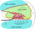
The cochlear duct is a fluid-filled cavity located within the cochlea. It is separated from the scala vestibuli by Reissner's membrane and from the scala tympani by the basilar membrane. The cochlear duct is filled with a fluid known as endolymph, which is different from the perilymph that fills the scala vestibuli and scala tympani.
Function[edit]
The primary function of the cochlear duct is to transmit sound vibrations from the environment to the auditory nerve. This is achieved through the movement of the endolymph within the cochlear duct, which stimulates the sensory cells of the organ of Corti. These cells then convert the mechanical vibrations into electrical signals that can be interpreted by the brain as sound.
Clinical Significance[edit]
Abnormalities or damage to the cochlear duct can result in various hearing disorders. For instance, Meniere's disease is a condition characterized by an excess of endolymph in the cochlear duct, leading to symptoms such as vertigo, tinnitus, and hearing loss.
See Also[edit]
References[edit]
<references group="" responsive="1"></references>

-
 Diagram of the internal ear anatomy
 -
 Cross-section of the cochlear duct
 -
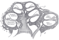
 Anatomy of the cochlea
 -
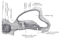
 Structure of the cochlear duct
 -
 Detailed view of the cochlear duct
 -
 Cross-section of the cochlea


