Vaginal ultrasonography: Difference between revisions
CSV import Tags: mobile edit mobile web edit |
CSV import |
||
| Line 39: | Line 39: | ||
[[Category:Ultrasound]] | [[Category:Ultrasound]] | ||
[[Category:Gynecology]] | [[Category:Gynecology]] | ||
<gallery> | |||
File:Transvaginal_ultrasonography_device.png|Transvaginal ultrasonography device | |||
File:Vaginal_Ultrasound.png|Vaginal ultrasound | |||
</gallery> | |||
Latest revision as of 01:45, 18 February 2025
Vaginal Ultrasonography[edit]

Vaginal ultrasonography is a type of pelvic ultrasound used by medical professionals to examine female reproductive organs. This includes the uterus, ovaries, fallopian tubes, cervix, and the vagina.
Procedure[edit]
Vaginal ultrasonography is performed using a transducer, which is a small, wand-like device that is inserted into the vagina. The transducer emits sound waves that bounce off the internal organs, creating images on a monitor. This procedure is often referred to as a transvaginal ultrasound.
The patient is usually asked to lie on her back with her feet in stirrups, similar to a gynecological examination. A protective cover is placed over the transducer, and a small amount of lubricant is applied to ease insertion. The procedure typically takes about 30 minutes and is generally painless, though some women may experience mild discomfort.
Uses[edit]
Vaginal ultrasonography is used for a variety of diagnostic purposes, including:
- Evaluating unexplained pelvic pain
- Investigating abnormal vaginal bleeding
- Monitoring ovarian cysts
- Assessing uterine fibroids
- Diagnosing ectopic pregnancy
- Monitoring fetal development in early pregnancy
Advantages[edit]
Compared to abdominal ultrasound, vaginal ultrasonography provides a clearer and more detailed view of the pelvic organs. This is because the transducer is closer to the organs being examined, allowing for higher resolution images. It is particularly useful in early pregnancy and for evaluating conditions that affect the uterus and ovaries.
Limitations[edit]
While vaginal ultrasonography is a valuable diagnostic tool, it has limitations. It may not be suitable for women who have never been sexually active or for those with certain medical conditions. Additionally, it may not provide sufficient information in cases where a broader view of the pelvic area is needed.
Related Pages[edit]
References[edit]
<references group="" responsive="1"></references>
-
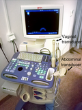 Transvaginal ultrasonography device
 -
 Vaginal ultrasound

