External vertebral venous plexuses
External Vertebral Venous Plexuses[edit]
The external vertebral venous plexuses are a network of veins that surround the vertebral column. They play a crucial role in the venous drainage of the spinal cord and surrounding structures. This article will provide a detailed overview of the external vertebral venous plexuses, including their anatomy, function, and clinical significance.
Anatomy[edit]
The external vertebral venous plexuses consist of a complex network of veins that are located outside the vertebral column. They are divided into two main plexuses: the anterior external vertebral venous plexus and the posterior external vertebral venous plexus.
The anterior external vertebral venous plexus is located in front of the vertebral bodies. It is formed by the union of veins from the spinal cord, intervertebral discs, and surrounding structures. These veins drain into the internal vertebral venous plexus, which is located within the vertebral canal.
The posterior external vertebral venous plexus is located behind the vertebral bodies. It is formed by the union of veins from the spinal cord, intervertebral discs, and surrounding structures. These veins drain into the internal vertebral venous plexus or the paravertebral venous plexus.
Function[edit]
The external vertebral venous plexuses play a crucial role in the venous drainage of the spinal cord and surrounding structures. They provide an alternative pathway for blood to drain from the spinal cord when the internal vertebral venous plexus is compromised.
The external vertebral venous plexuses also serve as a communication pathway between the veins of the spinal cord and the veins of the thorax and abdomen. This allows for the exchange of blood and nutrients between these regions.
Clinical Significance[edit]
The external vertebral venous plexuses have important clinical implications. They can serve as a potential route for the spread of cancer cells from the spine to other parts of the body. This is known as vertebral metastasis and can lead to the development of secondary tumors in distant organs.
In addition, the external vertebral venous plexuses can become engorged and dilated in certain conditions, such as spinal cord compression or trauma. This can result in venous congestion and increased pressure within the spinal cord, leading to symptoms such as pain, numbness, and weakness.
Internal Links[edit]
To learn more about related topics, please follow the internal links below:
- Vertebral column: Provides an overview of the structure and function of the vertebral column. - Venous drainage of the spinal cord: Explores the venous drainage system of the spinal cord. - Internal vertebral venous plexus: Discusses the anatomy and function of the internal vertebral venous plexus. - Paravertebral venous plexus: Provides information on the paravertebral venous plexus, another important venous drainage pathway in the spine.
References[edit]
Please add relevant references here.
-
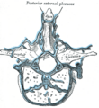 Gray's Anatomy illustration of the external vertebral venous plexuses

Medical Disclaimer: WikiMD is for informational purposes only and is not a substitute for professional medical advice. Content may be inaccurate or outdated and should not be used for diagnosis or treatment. Always consult your healthcare provider for medical decisions. Verify information with trusted sources such as CDC.gov and NIH.gov. By using this site, you agree that WikiMD is not liable for any outcomes related to its content. See full disclaimer.
Credits:Most images are courtesy of Wikimedia commons, and templates, categories Wikipedia, licensed under CC BY SA or similar.
Translate this page: - East Asian
中文,
日本,
한국어,
South Asian
हिन्दी,
தமிழ்,
తెలుగు,
Urdu,
ಕನ್ನಡ,
Southeast Asian
Indonesian,
Vietnamese,
Thai,
မြန်မာဘာသာ,
বাংলা
European
español,
Deutsch,
français,
Greek,
português do Brasil,
polski,
română,
русский,
Nederlands,
norsk,
svenska,
suomi,
Italian
Middle Eastern & African
عربى,
Turkish,
Persian,
Hebrew,
Afrikaans,
isiZulu,
Kiswahili,
Other
Bulgarian,
Hungarian,
Czech,
Swedish,
മലയാളം,
मराठी,
ਪੰਜਾਬੀ,
ગુજરાતી,
Portuguese,
Ukrainian
