Inclusion bodies
Inclusion bodies are distinct structures formed within the cells, often observed under the microscope in the cytoplasm or nucleus. These entities can be indicative of viral infections, genetic diseases, or other cellular stress conditions. Inclusion bodies are significant in both clinical diagnostics and research, providing insights into disease mechanisms and cellular responses to various stresses.
Formation and Types[edit]
Inclusion bodies form when proteins aggregate within a cell. This aggregation can be due to an overproduction of proteins, mutations that lead to misfolded proteins, or the presence of foreign proteins, such as those produced during viral infections. There are two main types of inclusion bodies: Nuclear inclusion bodies and Cytoplasmic inclusion bodies.
Nuclear Inclusion Bodies[edit]
Nuclear inclusion bodies are found within the nucleus of the cell. They are often associated with viral infections, where viral DNA or RNA accumulates, or with genetic disorders that lead to the abnormal accumulation of proteins. Examples include the Intranuclear inclusion bodies seen in cells infected with herpes simplex virus.
Cytoplasmic Inclusion Bodies[edit]
Cytoplasmic inclusion bodies appear in the cytoplasm and can be either pathological or non-pathological. Pathological inclusion bodies are typically associated with diseases, such as Lewy bodies in Parkinson's disease or Mallory bodies in alcoholic liver disease. Non-pathological inclusion bodies, such as Russell bodies and Glycogen bodies, may form as part of normal cellular processes.
Clinical Significance[edit]
Inclusion bodies are of significant clinical interest because their presence can help in diagnosing specific diseases. For example, the detection of Negri bodies is crucial for the diagnosis of rabies. Similarly, the presence of Lewy bodies is a key diagnostic marker for Parkinson's disease.
Research Applications[edit]
In biotechnology and protein engineering, inclusion bodies can be a challenge as they often contain recombinant proteins of interest in an insoluble and inactive form. However, recent advances have enabled the recovery of functional proteins from inclusion bodies, providing a valuable source of proteins for research and therapeutic applications.
Management and Treatment[edit]
The presence of inclusion bodies in diseases often necessitates a management strategy focused on the underlying condition. For example, in viral infections, antiviral therapies may reduce the formation of viral inclusion bodies. In genetic disorders, strategies may include gene therapy or the use of molecular chaperones to prevent protein misfolding.
Conclusion[edit]
Inclusion bodies serve as a window into the cellular response to stress, infection, and genetic mutations. Their study not only aids in the diagnosis and understanding of various diseases but also offers potential avenues for therapeutic intervention.
This medical article is a stub. You can help WikiMD by expanding the page. |
-
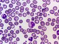 Canine Distemper Virus Cytoplasmic Inclusion Body (Blood smear, Wright's stain)
.jpg)

Medical Disclaimer: WikiMD is for informational purposes only and is not a substitute for professional medical advice. Content may be inaccurate or outdated and should not be used for diagnosis or treatment. Always consult your healthcare provider for medical decisions. Verify information with trusted sources such as CDC.gov and NIH.gov. By using this site, you agree that WikiMD is not liable for any outcomes related to its content. See full disclaimer.
Credits:Most images are courtesy of Wikimedia commons, and templates, categories Wikipedia, licensed under CC BY SA or similar.
Translate page: - East Asian
中文,
日本,
한국어,
South Asian
हिन्दी,
தமிழ்,
తెలుగు,
Urdu,
ಕನ್ನಡ,
Southeast Asian
Indonesian,
Vietnamese,
Thai,
မြန်မာဘာသာ,
বাংলা
European
español,
Deutsch,
français,
Greek,
português do Brasil,
polski,
română,
русский,
Nederlands,
norsk,
svenska,
suomi,
Italian
Middle Eastern & African
عربى,
Turkish,
Persian,
Hebrew,
Afrikaans,
isiZulu,
Kiswahili,
Other
Bulgarian,
Hungarian,
Czech,
Swedish,
മലയാളം,
मराठी,
ਪੰਜਾਬੀ,
ગુજરાતી,
Portuguese,
Ukrainian
