File:Ultrastructure of tracheal hemidesmosomes in mice.JPEG
From WikiMD's medical encyclopedia

Size of this preview: 497 × 600 pixels. Other resolutions: 199 × 240 pixels | 398 × 480 pixels | 994 × 1,200 pixels.
Original file (994 × 1,200 pixels, file size: 350 KB, MIME type: image/jpeg)
| Description |
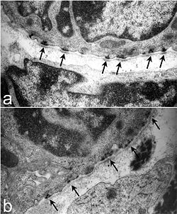
English: Ultrastructure of tracheal hemidesmosomes. Tracheas from P1-2 Lamc2-/- and littermate control mice were processed for transmission electron microscopy. Littermate control tracheas (a) had well-defined, organized hemidesmosomes with darkened areas in the lamina densa abutting the hemidesmosome (arrows). In contrast, hemidesmosomes in Lamc2-/- tracheas (b) were less organized, the intracellular component was more diffuse, and the lamina densa directly below the hemidesmosomal areas lacked the electron density seen in the littermate control (arrows). Nguyen et al. Respiratory Research 2006 7:28 doi:10.1186/1465-9921-7-28 |
| Date | |
| Source | Lung development in laminin γ2 deficiency: abnormal tracheal hemidesmosomes with normal branching morphogenesis and epithelial differentiation |
| Author | Nguyen NM, Pulkkinen L, Schlueter JA, Meneguzzi G, Uitto J, Senior RM. |
| Permission (Reusing this file) |
© 2006 Nguyen et al; licensee BioMed Central Ltd. This is an Open Access article distributed under the terms of the Creative Commons Attribution License (https://creativecommons.org/licenses/by/2.0), which permits unrestricted use, distribution, and reproduction in any medium, provided the original work is properly cited. |
This file is licensed under the Creative Commons Attribution-Share Alike 2.0 Generic license.
- You are free:
- to share – to copy, distribute and transmit the work
- to remix – to adapt the work
- Under the following conditions:
- attribution – You must give appropriate credit, provide a link to the license, and indicate if changes were made. You may do so in any reasonable manner, but not in any way that suggests the licensor endorses you or your use.
- share alike – If you remix, transform, or build upon the material, you must distribute your contributions under the same or compatible license as the original.
File history
Click on a date/time to view the file as it appeared at that time.
| Date/Time | Thumbnail | Dimensions | User | Comment | |
|---|---|---|---|---|---|
| current | 10:00, 10 July 2009 | | 994 × 1,200 (350 KB) | CopperKettle | {{Information |Description={{en|1=Ultrastructure of tracheal hemidesmosomes. Tracheas from P1-2 Lamc2-/- and littermate control mice were processed for transmission electron microscopy. Littermate control tracheas (a) had well-defined, organized hemidesmo |
File usage
The following 2 pages use this file: