File:Pulmonary embolism.jpg
From WikiMD's medical encyclopedia

Size of this preview: 800 × 581 pixels. Other resolutions: 320 × 233 pixels | 640 × 465 pixels | 922 × 670 pixels.
Original file (922 × 670 pixels, file size: 55 KB, MIME type: image/jpeg)
| Description |
Čeština: CT - plicní embolie
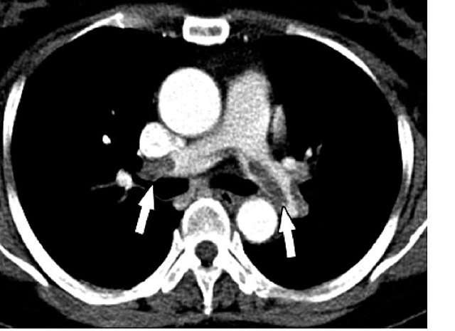
English: Chest Spiral CT (with and without contrast agent) showing multiples filling defects of principal branches, due to acute and chronic pulmonary embolism. |
| Date | Published: 24 August 2007 |
| Source | Pulmonary embolism and patent foramen ovale thrombosis: the key role of TEE. Cardiovascular Ultrasound 2007, 5:26. doi:10.1186/1476-7120-5-26 |
| Author | Walter Serra, Giuseppe De Iaco, Claudio Reverberi and Tiziano Gherli |
| Permission (Reusing this file) |
This file is licensed under the Creative Commons Attribution 2.0 Generic license.
|
File history
Click on a date/time to view the file as it appeared at that time.
| Date/Time | Thumbnail | Dimensions | User | Comment | |
|---|---|---|---|---|---|
| current | 16:09, 29 December 2008 | | 922 × 670 (55 KB) | Stevenfruitsmaak | {{Information |Description={{en|1=Chest Spiral CT (with and without contrast agent) showing multiples filling defects of principal branches, due to acute and chronic pulmonary embolism.}} |Source=[http://www.cardiovascularultrasound.com/content/5/1/26 Pul |
File usage
The following page uses this file: