File:Perinexial ephaptic coupling.jpg
From WikiMD's medical encyclopedia
Size of this preview: 470 × 599 pixels. Other resolutions: 188 × 240 pixels | 546 × 696 pixels.
Original file (546 × 696 pixels, file size: 106 KB, MIME type: image/jpeg)
Summary
| Description |
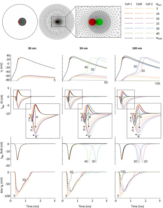
English: Top: schematic and mesh of the ID (inset showing its high density region), with the central Na+ channel cluster (red) located inside the perinexus (grey) of a closely apposed gap junction plaque (green). Simulation results for G gap = 0 nS and bulk cleft widths w cleft of 30 nm (left column), 50 nm (middle column) and 100 nm (right column). Perinexus width w peri was varied from 5 nm up to the bulk cleft width (see colour key upper right). First row: intracellular potentialAn external file that holds a picture, illustration, etc. Object name is TJP-599-4779-e028.jpgin cell 1 (continuous lines) and cell 2 (dashed lines); second and third rows: Na+ current in the ID and the bulk membrane of cell 1 (continuous lines) and cell 2 (dashed lines), respectively; fourth row: minimal extracellular potential in the cleft (dash‐dotted lines) as a function of time. The labels of selected traces indicate w peri. The rectangles denote the portions of the plots that were magnified (insets). |
| Date | |
| Source | J Physiol. 2021 Nov;599(21):4779-4811. doi: 10.1113/JP282105. Epub 2021 Oct 4. |
| Author | Ena Ivanovic, Jan P Kucera |
Licensing
This file is licensed under the Creative Commons Attribution-Share Alike 4.0 International license.
- You are free:
- to share – to copy, distribute and transmit the work
- to remix – to adapt the work
- Under the following conditions:
- attribution – You must give appropriate credit, provide a link to the license, and indicate if changes were made. You may do so in any reasonable manner, but not in any way that suggests the licensor endorses you or your use.
- share alike – If you remix, transform, or build upon the material, you must distribute your contributions under the same or compatible license as the original.
File history
Click on a date/time to view the file as it appeared at that time.
| Date/Time | Thumbnail | Dimensions | User | Comment | |
|---|---|---|---|---|---|
| current | 03:53, 28 May 2023 | | 546 × 696 (106 KB) | Tgru001 | Uploaded a work by Ena Ivanovic, Jan P Kucera from J Physiol. 2021 Nov;599(21):4779-4811. doi: 10.1113/JP282105. Epub 2021 Oct 4. with UploadWizard |
File usage
The following page uses this file: