File:Mitosis Stages.svg
From WikiMD's medical encyclopedia

Size of this PNG preview of this SVG file: 800 × 139 pixels. Other resolutions: 320 × 55 pixels | 640 × 111 pixels | 1,024 × 177 pixels | 1,280 × 222 pixels | 2,560 × 443 pixels | 2,361 × 409 pixels.
Original file (SVG file, nominally 2,361 × 409 pixels, file size: 1.13 MB)
Summary
| Description |
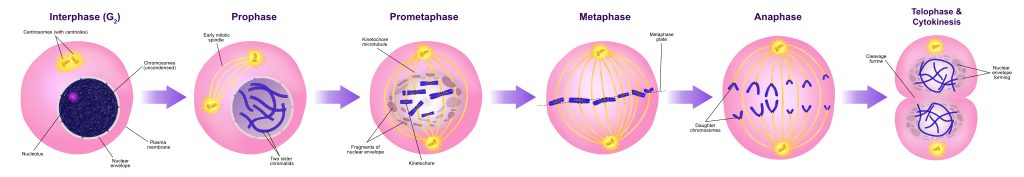
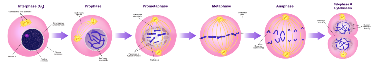
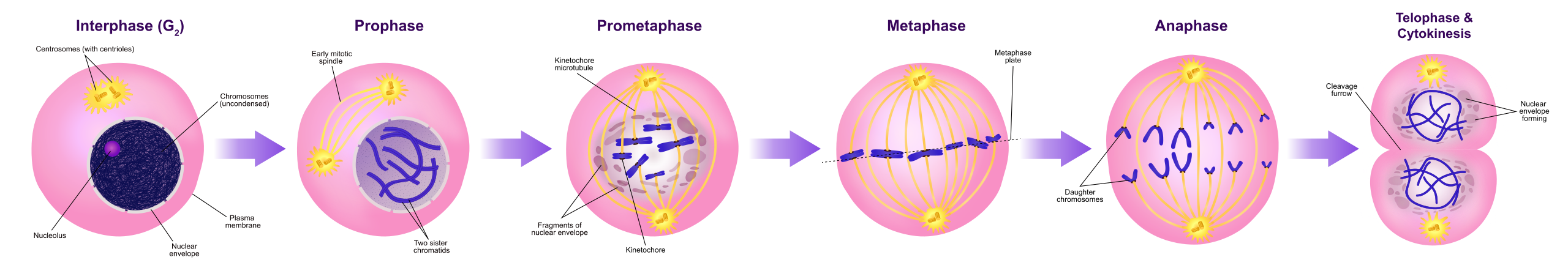
English: A diagram of mitosis stages Interphase (G₂): In this substage, the cell prepares for nuclear division and a protein that makes microtubles for cell division is synthesized. Prophase: The longest stage of mitosis. In this stage the chromosomes become visible and the centrioles separate and move to opposite poles of the cell. Prometaphase: The nuclear envelope disintegrates and microtubules can attach to kinetochores. Chromosomes congress toward the metaphase plate of the cell. Metaphase: In this stage the chromosomes line up across the center of the cell and become connected to the spindle fiber at their centromere. Anaphase: In this stage the sister chromatids separate into individual chromosomes and are pulled apart. Telophase & cytokinesis: Chromosomes decondense and are surrounded by a newly formed nuclear envelope. Cytokinesis typically coincides with and telophase. |
||
| Date | |||
| Source |
Own work; Used information from: Campbell Biology (10th Edition) by: Jane B. Reece & Steven A. Wasserman. and Nature.com. |
||
| Author | Ali Zifan | ||
| Other versions |
[]
|
||
| SVG development |
|
|
Licensing
I, the copyright holder of this work, hereby publish it under the following license:
This file is licensed under the Creative Commons Attribution-Share Alike 4.0 International license.
- You are free:
- to share – to copy, distribute and transmit the work
- to remix – to adapt the work
- Under the following conditions:
- attribution – You must give appropriate credit, provide a link to the license, and indicate if changes were made. You may do so in any reasonable manner, but not in any way that suggests the licensor endorses you or your use.
- share alike – If you remix, transform, or build upon the material, you must distribute your contributions under the same or compatible license as the original.

|
This image has been assessed under the valued image criteria and is considered the most valued image on Commons within the scope: Biology diagrams, Cell diagrams, and Mitosis. You can see its nomination here. |
File history
Click on a date/time to view the file as it appeared at that time.
| Date/Time | Thumbnail | Dimensions | User | Comment | |
|---|---|---|---|---|---|
| current | 01:30, 1 March 2021 | 2,361 × 409 (1.13 MB) | Cmglee | Fix misspelling of "mitotic" by cloning path5986 as path5983. |
File usage
The following page uses this file: