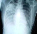
Chest radiograph
Chest Radiograph[edit]
A chest radiograph, commonly known as a chest X-ray, is a medical imaging technique used to visualize the internal structures of the chest. It is a non-invasive and widely available diagnostic tool that provides valuable information about the heart, lungs, and other structures within the chest cavity.
Procedure[edit]
During a chest radiograph, the patient stands in front of a specialized X-ray machine. The technician positions the patient's chest against the machine's detector plate and instructs them to take a deep breath and hold it. This helps to obtain a clear image of the chest.
The X-ray machine emits a small amount of radiation, which passes through the patient's chest and is captured by the detector plate. The resulting image is then processed and displayed on a computer screen for further analysis.
Uses[edit]
Chest radiographs are commonly used to diagnose and monitor various medical conditions. They provide valuable information about the size, shape, and position of the heart, lungs, and other structures within the chest cavity. Some of the common uses of chest radiographs include:
1. **Diagnosing lung diseases**: Chest radiographs can help identify conditions such as pneumonia, tuberculosis, lung cancer, and chronic obstructive pulmonary disease (COPD).
2. **Evaluating heart health**: Chest radiographs can reveal the size and shape of the heart, as well as detect abnormalities such as an enlarged heart or fluid accumulation around the heart.
3. **Assessing chest injuries**: Chest radiographs are often used to evaluate the extent of chest injuries, such as rib fractures or lung contusions, in cases of trauma or accidents.
4. **Monitoring treatment progress**: Chest radiographs can be used to monitor the effectiveness of treatments for various conditions, such as pneumonia or lung cancer.
Interpretation[edit]
Interpreting a chest radiograph requires specialized knowledge and expertise. Radiologists, who are medical doctors with specialized training in medical imaging, are responsible for analyzing and interpreting the images.
When interpreting a chest radiograph, radiologists look for various signs and abnormalities, including:
1. **Lung abnormalities**: These can include areas of consolidation (indicative of pneumonia), nodules or masses (suggestive of lung cancer), or signs of chronic lung diseases.
2. **Heart abnormalities**: Radiologists assess the size, shape, and position of the heart, looking for signs of enlargement, fluid accumulation, or structural abnormalities.
3. **Bone abnormalities**: Chest radiographs can also reveal fractures, dislocations, or other bone-related abnormalities in the chest area.
Conclusion[edit]
Chest radiographs are a valuable diagnostic tool in the field of medicine. They provide important information about the heart, lungs, and other structures within the chest cavity, aiding in the diagnosis and monitoring of various medical conditions. With the expertise of radiologists, chest radiographs play a crucial role in providing accurate and timely medical diagnoses.
See Also[edit]
References[edit]
<references />
-
Normal posteroanterior (PA) chest radiograph (X-ray)
-
Dedicated chest x-ray room
-
Hospital Corpsman taking a chest X-ray
-
Normal lateral chest radiograph (X-ray)
-
Chest labeled
-
Mediastinal structures on chest X-ray, annotated
-
Pneumonia wedge
-
X-ray of an infant with a prominent thymus
-
Supraclavicular fossa on chest X-ray
-
Chest Xray PA inverted
-
Projectional rendering of CT scan of thorax
-
SARS xray
Ad. Transform your life with W8MD's Budget GLP-1 injections from $75


W8MD offers a medical weight loss program to lose weight in Philadelphia. Our physician-supervised medical weight loss provides:
- Weight loss injections in NYC (generic and brand names):
- Zepbound / Mounjaro, Wegovy / Ozempic, Saxenda
- Most insurances accepted or discounted self-pay rates. We will obtain insurance prior authorizations if needed.
- Generic GLP1 weight loss injections from $75 for the starting dose.
- Also offer prescription weight loss medications including Phentermine, Qsymia, Diethylpropion, Contrave etc.
NYC weight loss doctor appointmentsNYC weight loss doctor appointments
Start your NYC weight loss journey today at our NYC medical weight loss and Philadelphia medical weight loss clinics.
- Call 718-946-5500 to lose weight in NYC or for medical weight loss in Philadelphia 215-676-2334.
- Tags:NYC medical weight loss, Philadelphia lose weight Zepbound NYC, Budget GLP1 weight loss injections, Wegovy Philadelphia, Wegovy NYC, Philadelphia medical weight loss, Brookly weight loss and Wegovy NYC
|
WikiMD's Wellness Encyclopedia |
| Let Food Be Thy Medicine Medicine Thy Food - Hippocrates |
Medical Disclaimer: WikiMD is not a substitute for professional medical advice. The information on WikiMD is provided as an information resource only, may be incorrect, outdated or misleading, and is not to be used or relied on for any diagnostic or treatment purposes. Please consult your health care provider before making any healthcare decisions or for guidance about a specific medical condition. WikiMD expressly disclaims responsibility, and shall have no liability, for any damages, loss, injury, or liability whatsoever suffered as a result of your reliance on the information contained in this site. By visiting this site you agree to the foregoing terms and conditions, which may from time to time be changed or supplemented by WikiMD. If you do not agree to the foregoing terms and conditions, you should not enter or use this site. See full disclaimer.
Credits:Most images are courtesy of Wikimedia commons, and templates, categories Wikipedia, licensed under CC BY SA or similar.
Translate this page: - East Asian
中文,
日本,
한국어,
South Asian
हिन्दी,
தமிழ்,
తెలుగు,
Urdu,
ಕನ್ನಡ,
Southeast Asian
Indonesian,
Vietnamese,
Thai,
မြန်မာဘာသာ,
বাংলা
European
español,
Deutsch,
français,
Greek,
português do Brasil,
polski,
română,
русский,
Nederlands,
norsk,
svenska,
suomi,
Italian
Middle Eastern & African
عربى,
Turkish,
Persian,
Hebrew,
Afrikaans,
isiZulu,
Kiswahili,
Other
Bulgarian,
Hungarian,
Czech,
Swedish,
മലയാളം,
मराठी,
ਪੰਜਾਬੀ,
ગુજરાતી,
Portuguese,
Ukrainian


