Cone dystrophy
Cone dystrophy is a group of rare eye disorders that affect the cone cells, the cells responsible for color vision and visual acuity. Cone dystrophies can cause a variety of symptoms such as decreased visual acuity, color blindness, increased sensitivity to light, and an abnormal sensitivity to light (photophobia).
Symptoms
The symptoms of cone dystrophy can vary greatly from person to person, even among members of the same family. The most common symptoms include:
- Decreased visual acuity: This is often the first symptom of cone dystrophy. It usually begins in childhood or adolescence and gradually worsens over time.
- Color blindness: This is a common symptom of cone dystrophy. It can range from mild to severe, and it often affects the ability to distinguish between certain colors.
- Photophobia: This is an abnormal sensitivity to light. It can make it difficult to be in bright environments and can cause discomfort or pain in the eyes.
- Loss of central vision: This can occur in the later stages of cone dystrophy. It can make it difficult to read, drive, or recognize faces.
Causes
Cone dystrophy is usually caused by genetic mutations. It is most often inherited in an autosomal dominant manner, which means one copy of the altered gene in each cell is sufficient to cause the disorder. However, it can also be inherited in an autosomal recessive manner, which means both copies of the gene in each cell have mutations.
Diagnosis
Diagnosis of cone dystrophy is based on the clinical symptoms, a detailed patient history, and specialized tests that examine the functioning of the cone cells. These tests may include an electroretinogram (ERG), which measures the electrical responses of the light-sensitive cells in the eyes.
Treatment
There is currently no cure for cone dystrophy. Treatment is aimed at managing the symptoms and improving the quality of life. This may include the use of low-vision aids, such as magnifying devices, and the use of sunglasses to help with light sensitivity.
See also
References
This medical article is a stub. You can help WikiMD by expanding the page. |
-
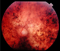 Fundus of a patient with cone-rod dystrophy

Medical Disclaimer: WikiMD is for informational purposes only and is not a substitute for professional medical advice. Content may be inaccurate or outdated and should not be used for diagnosis or treatment. Always consult your healthcare provider for medical decisions. Verify information with trusted sources such as CDC.gov and NIH.gov. By using this site, you agree that WikiMD is not liable for any outcomes related to its content. See full disclaimer.
Credits:Most images are courtesy of Wikimedia commons, and templates, categories Wikipedia, licensed under CC BY SA or similar.
Translate this page: - East Asian
中文,
日本,
한국어,
South Asian
हिन्दी,
தமிழ்,
తెలుగు,
Urdu,
ಕನ್ನಡ,
Southeast Asian
Indonesian,
Vietnamese,
Thai,
မြန်မာဘာသာ,
বাংলা
European
español,
Deutsch,
français,
Greek,
português do Brasil,
polski,
română,
русский,
Nederlands,
norsk,
svenska,
suomi,
Italian
Middle Eastern & African
عربى,
Turkish,
Persian,
Hebrew,
Afrikaans,
isiZulu,
Kiswahili,
Other
Bulgarian,
Hungarian,
Czech,
Swedish,
മലയാളം,
मराठी,
ਪੰਜਾਬੀ,
ગુજરાતી,
Portuguese,
Ukrainian
