Scanning laser ophthalmoscopy
Scanning Laser Ophthalmoscopy (SLO) is a method of imaging the retina that has revolutionized the way ophthalmology and optometry examine the back of the eye. Unlike traditional fundus photography, which uses a flash to illuminate the retina, SLO employs a low-power laser beam that scans the retina in a raster pattern. This technique allows for detailed, high-contrast images of the retinal structure, providing invaluable information for the diagnosis and management of various eye diseases.
Overview
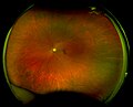
Scanning Laser Ophthalmoscopy utilizes a laser as its light source to illuminate the retina. The reflected light from the retina is then detected by a sensitive camera. Because the laser scans the retina point by point, it can construct a highly detailed image of the retinal surface. This method reduces the scatter of light and allows for imaging through media opacities such as cataracts, which can obscure traditional imaging methods.
Advantages
The primary advantages of SLO include its ability to provide high-resolution images of the retina, its utility in imaging through opacities, and its capacity to perform various types of functional imaging. For instance, SLO can be adapted to perform angiography without the need for dye (non-invasive), autofluorescence imaging to assess retinal health, and optical coherence tomography (OCT) for cross-sectional images of the retina.
Applications
SLO is used in the diagnosis and management of a wide range of retinal conditions, including age-related macular degeneration (AMD), diabetic retinopathy, and glaucoma. It is particularly useful in tracking disease progression and response to treatment. Additionally, SLO can be used in research settings to study retinal diseases at a microscopic level.
Limitations
While SLO offers many advantages, it also has some limitations. The quality of the images can be affected by patient movement, and the technique requires cooperation from the patient to fixate on a target for a period of time. Additionally, the cost of SLO equipment can be prohibitive for some practices.
Future Directions
Advancements in SLO technology continue to expand its applications and improve image quality. Innovations such as adaptive optics SLO (AO-SLO) are enhancing the resolution of SLO images to a cellular level, allowing for detailed visualization of individual photoreceptor cells. This could lead to earlier detection and better understanding of retinal diseases.

-
 Scanning laser ophthalmoscopy
 -
 Scanning laser ophthalmoscopy


Ad. Transform your health with W8MD Weight Loss, Sleep & MedSpa

Tired of being overweight?
Get started with evidence based, physician-supervised
affordable GLP-1 weight loss injections
Now available in New York City and Philadelphia:
- Semaglutide starting from $59.99/week and up
- Tirzepatide starting from $69.99/week and up (dose dependent)
✔ Evidence-based medical weight loss ✔ Insurance-friendly visits available ✔ Same-week appointments, evenings & weekends
Learn more:
Start your transformation today with W8MD weight loss centers.
|
WikiMD Medical Encyclopedia |
Medical Disclaimer: WikiMD is for informational purposes only and is not a substitute for professional medical advice. Content may be inaccurate or outdated and should not be used for diagnosis or treatment. Always consult your healthcare provider for medical decisions. Verify information with trusted sources such as CDC.gov and NIH.gov. By using this site, you agree that WikiMD is not liable for any outcomes related to its content. See full disclaimer.
Credits:Most images are courtesy of Wikimedia commons, and templates, categories Wikipedia, licensed under CC BY SA or similar.
Translate this page: - East Asian
中文,
日本,
한국어,
South Asian
हिन्दी,
தமிழ்,
తెలుగు,
Urdu,
ಕನ್ನಡ,
Southeast Asian
Indonesian,
Vietnamese,
Thai,
မြန်မာဘာသာ,
বাংলা
European
español,
Deutsch,
français,
Greek,
português do Brasil,
polski,
română,
русский,
Nederlands,
norsk,
svenska,
suomi,
Italian
Middle Eastern & African
عربى,
Turkish,
Persian,
Hebrew,
Afrikaans,
isiZulu,
Kiswahili,
Other
Bulgarian,
Hungarian,
Czech,
Swedish,
മലയാളം,
मराठी,
ਪੰਜਾਬੀ,
ગુજરાતી,
Portuguese,
Ukrainian
