Scanning helium ion microscope: Difference between revisions
CSV import |
CSV import |
||
| Line 33: | Line 33: | ||
{{Microscopy-stub}} | {{Microscopy-stub}} | ||
== Scanning helium ion microscope gallery == | |||
<gallery> | |||
File:ORION NanoFab - Helium Ion Microscope (8410606251).jpg|ORION NanoFab - Helium Ion Microscope | |||
File:SEM vs HIM imaging of mouse enamel.jpg|SEM vs HIM imaging of mouse enamel | |||
</gallery> | |||
Latest revision as of 05:35, 3 March 2025
Scanning Helium Ion Microscope (SHIM) is a type of microscope that uses a focused beam of helium ions to obtain high-resolution images of samples. Unlike traditional electron microscopy, which uses electrons for imaging, SHIM offers several advantages, including higher surface detail resolution and minimal sample damage. This technology has become increasingly important in fields such as material science, biology, and nanotechnology due to its ability to provide detailed surface images and its capacity for material analysis.
Overview[edit]
The Scanning Helium Ion Microscope operates by focusing a beam of helium ions onto a sample and detecting the secondary electrons that are emitted from the sample surface. The helium ion beam is generated in an ion source, typically using a gas field ion source (GFIS), and then accelerated towards the sample. The interaction of the helium ions with the sample surface results in the emission of secondary electrons, which are collected to form an image.
Advantages[edit]
SHIM offers several advantages over traditional electron microscopy techniques:
- Higher Resolution: Due to the smaller wavelength of helium ions compared to electrons, SHIM can achieve higher resolution images, allowing for the observation of finer details on the sample surface.
- Reduced Sample Damage: Helium ions penetrate less deeply into the sample than electrons, resulting in less damage and allowing for the imaging of delicate materials.
- Charge Compensation: Helium ion microscopy can be used on insulating materials without the need for conductive coatings, as the helium ions can neutralize the charge build-up on the sample surface.
- Material Contrast: SHIM provides enhanced material contrast, making it easier to differentiate between different materials in a sample.
Applications[edit]
The unique capabilities of SHIM have led to its application in a variety of fields:
- In material science, it is used for the characterization of nanomaterials, thin films, and semiconductors.
- In biology, SHIM is utilized for imaging biological samples at high resolution, including cells and tissues, without the need for staining or extensive sample preparation.
- In nanotechnology, it aids in the development and inspection of nanoscale devices and structures.
Limitations[edit]
While SHIM offers many advantages, there are also some limitations to consider:
- Cost: The cost of a Scanning Helium Ion Microscope is higher than that of traditional electron microscopes, limiting its accessibility.
- Sample Preparation: Although less extensive than for electron microscopy, some sample preparation is still required to ensure optimal imaging results.
- Field of View: The field of view in SHIM is generally smaller than that in electron microscopy, which can be a limitation for certain applications.
Future Directions[edit]
Research and development in the field of helium ion microscopy continue to expand its capabilities and applications. Future advancements may include improved ion sources for even higher resolution imaging, enhanced analytical capabilities for material characterization, and increased accessibility of the technology for a wider range of research fields.
This article is a Microscopy-related stub. You can help WikiMD by expanding it!
Scanning helium ion microscope gallery[edit]
-
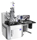 ORION NanoFab - Helium Ion Microscope
 -
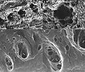 SEM vs HIM imaging of mouse enamel
.jpg)

