Radiographic anatomy: Difference between revisions
CSV import |
CSV import |
||
| Line 48: | Line 48: | ||
{{Radiology-stub}} | {{Radiology-stub}} | ||
== Radiographic_anatomy == | |||
<gallery> | |||
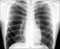
File:Chest.jpg|Chest | |||
File:Chestxp_illustrated.jpg|Chestxp illustrated | |||
</gallery> | |||
Latest revision as of 21:38, 23 February 2025
Radiographic Anatomy refers to the study of the human body as it appears on radiographic images. This field is essential for medical professionals, particularly radiologists, radiographers, and anatomists, who rely on these images to diagnose, treat, and understand human anatomy and pathologies. Radiographic anatomy encompasses various imaging modalities, including X-ray, Computed Tomography (CT), Magnetic Resonance Imaging (MRI), and Ultrasound.
Overview[edit]
Radiographic images provide a two-dimensional representation of the three-dimensional human body. Understanding these images requires a thorough knowledge of human anatomy and the principles of radiographic imaging. The contrast in radiographic images is generated by the differential absorption of X-rays by different tissues, with denser tissues such as bone appearing white, and less dense tissues such as muscle and fat appearing in shades of gray.
Importance[edit]
The importance of radiographic anatomy lies in its utility for medical diagnosis and treatment. It allows healthcare professionals to:
- Identify normal anatomical structures and variations.
- Detect abnormalities such as fractures, tumors, and infections.
- Guide surgical procedures and treatments.
- Monitor the progression of diseases and the effectiveness of treatments.
Key Anatomical Regions[edit]
Radiographic anatomy is divided into several key regions, each with its own set of structures visible on radiographic images:
Chest[edit]
The chest or thoracic region includes the lungs, heart, ribs, and diaphragm. Common radiographic examinations of the chest are used to diagnose conditions such as pneumonia, heart failure, and lung cancer.
Abdomen[edit]
The abdomen contains organs of the digestive system, urinary system, and major blood vessels. Abdominal X-rays can reveal issues like bowel obstructions, kidney stones, and abdominal aortic aneurysms.
Skull and Brain[edit]
Skull and brain imaging, particularly through CT and MRI, is crucial for identifying fractures, hemorrhages, tumors, and neurological conditions.
Spine[edit]
The spine is composed of vertebrae, intervertebral discs, and the spinal cord. Radiographic imaging of the spine can detect scoliosis, disc herniation, and spinal tumors.
Extremities[edit]
The extremities include the arms, legs, hands, and feet. Radiographs of these areas are commonly used to diagnose fractures, dislocations, and degenerative joint diseases.
Imaging Modalities[edit]
Different imaging modalities are used in radiographic anatomy, each with its own applications and advantages:
- X-ray: The most common form of radiographic imaging, useful for examining bones and certain soft tissues.
- CT: Provides detailed cross-sectional images of the body, ideal for diagnosing complex fractures and internal organ diseases.
- MRI: Offers superior soft tissue contrast, making it invaluable for imaging the brain, spinal cord, and musculoskeletal system.
- Ultrasound: Uses sound waves to produce images of soft tissues, often used in obstetrics and to image the abdominal organs.
Challenges and Considerations[edit]
Radiographic anatomy requires careful consideration of several factors, including radiation exposure, image quality, and patient positioning. Healthcare professionals must balance the need for diagnostic information with the risks associated with radiation exposure, particularly in sensitive populations such as children and pregnant women.
Conclusion[edit]
Radiographic anatomy is a cornerstone of modern medicine, providing a window into the human body that is crucial for diagnosis and treatment. As imaging technologies advance, the field continues to evolve, offering ever greater insights into human anatomy and pathology.
This radiology related article is a stub. You can help WikiMD by expanding it.
Radiographic_anatomy[edit]
-
 Chest
Chest -
 Chestxp illustrated


