Scanning laser ophthalmoscopy: Difference between revisions
CSV import Tags: mobile edit mobile web edit |
CSV import Tags: mobile edit mobile web edit |
||
| Line 20: | Line 20: | ||
{{medicine-stub}} | {{medicine-stub}} | ||
<gallery> | |||
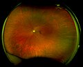
File:Healthy Adult OS, Color - California Projection.jpeg|Scanning laser ophthalmoscopy | |||
File:AOSLO setup labeled.png|Scanning laser ophthalmoscopy | |||
</gallery> | |||
Latest revision as of 01:43, 20 February 2025
Scanning Laser Ophthalmoscopy (SLO) is a method of imaging the retina that has revolutionized the way ophthalmology and optometry examine the back of the eye. Unlike traditional fundus photography, which uses a flash to illuminate the retina, SLO employs a low-power laser beam that scans the retina in a raster pattern. This technique allows for detailed, high-contrast images of the retinal structure, providing invaluable information for the diagnosis and management of various eye diseases.
Overview[edit]
Scanning Laser Ophthalmoscopy utilizes a laser as its light source to illuminate the retina. The reflected light from the retina is then detected by a sensitive camera. Because the laser scans the retina point by point, it can construct a highly detailed image of the retinal surface. This method reduces the scatter of light and allows for imaging through media opacities such as cataracts, which can obscure traditional imaging methods.
Advantages[edit]
The primary advantages of SLO include its ability to provide high-resolution images of the retina, its utility in imaging through opacities, and its capacity to perform various types of functional imaging. For instance, SLO can be adapted to perform angiography without the need for dye (non-invasive), autofluorescence imaging to assess retinal health, and optical coherence tomography (OCT) for cross-sectional images of the retina.
Applications[edit]
SLO is used in the diagnosis and management of a wide range of retinal conditions, including age-related macular degeneration (AMD), diabetic retinopathy, and glaucoma. It is particularly useful in tracking disease progression and response to treatment. Additionally, SLO can be used in research settings to study retinal diseases at a microscopic level.
Limitations[edit]
While SLO offers many advantages, it also has some limitations. The quality of the images can be affected by patient movement, and the technique requires cooperation from the patient to fixate on a target for a period of time. Additionally, the cost of SLO equipment can be prohibitive for some practices.
Future Directions[edit]
Advancements in SLO technology continue to expand its applications and improve image quality. Innovations such as adaptive optics SLO (AO-SLO) are enhancing the resolution of SLO images to a cellular level, allowing for detailed visualization of individual photoreceptor cells. This could lead to earlier detection and better understanding of retinal diseases.

-
 Scanning laser ophthalmoscopy
 -
 Scanning laser ophthalmoscopy


