Mitral regurgitation: Difference between revisions
CSV import |
CSV import |
||
| Line 52: | Line 52: | ||
[[Category:Cardiology]] | [[Category:Cardiology]] | ||
[[Category:Heart diseases]] | [[Category:Heart diseases]] | ||
<gallery> | |||
File:Mitral Regurgitation scheme1.png|Mitral Regurgitation Scheme | |||
File:Phonocardiograms from normal and abnormal heart sounds.svg|Phonocardiograms from Normal and Abnormal Heart Sounds | |||
File:Blausen 0645 MitralValve RegurgitationvsStenosis.png|Mitral Valve Regurgitation vs Stenosis | |||
File:MI Schema leicht Kopie.png|MI Schema | |||
File:Mitralinsuff TEE.jpg|Mitralinsuff TEE | |||
File:Mitral regurgitation echo 4chamber.jpg|Mitral Regurgitation Echo 4 Chamber | |||
File:Mitral regurgitation echo 4chamber description.png|Mitral Regurgitation Echo 4 Chamber Description | |||
File:Doppler mitral valve.gif|Doppler Mitral Valve | |||
File:The PISA Method for Quantification of Mitral Regurgitation.svg|The PISA Method for Quantification of Mitral Regurgitation | |||
</gallery> | |||
Revision as of 01:16, 20 February 2025
A condition where the heart's mitral valve does not close tightly, allowing blood to flow backward in the heart.
Overview
Mitral regurgitation (MR), also known as mitral insufficiency, is a disorder of the heart in which the mitral valve does not close properly when the heart pumps out blood. This improper closure allows blood to flow backward from the left ventricle into the left atrium, which can lead to various symptoms and complications.
Anatomy and Physiology
The mitral valve is one of the four valves in the heart, located between the left atrium and the left ventricle. It consists of two leaflets that open to allow blood to flow from the left atrium to the left ventricle and close to prevent backflow during ventricular contraction. Proper functioning of the mitral valve is crucial for maintaining efficient blood circulation.
Causes
Mitral regurgitation can be caused by a variety of factors, including:
- Mitral valve prolapse: A condition where the valve leaflets bulge into the left atrium during contraction.
- Rheumatic heart disease: Damage to the valve from rheumatic fever.
- Endocarditis: Infection of the heart valves.
- Ischemic heart disease: Damage to the heart muscle affecting valve function.
- Cardiomyopathy: Disease of the heart muscle that can affect valve function.
- Congenital heart defects: Birth defects affecting the structure of the heart.
Symptoms
Symptoms of mitral regurgitation can vary depending on the severity of the condition. They may include:
- Shortness of breath, especially during exertion or when lying flat
- Fatigue
- Palpitations
- Swelling in the legs or feet
- Cough, especially at night or when lying down
Diagnosis
Mitral regurgitation is typically diagnosed using:
- Echocardiography: An ultrasound of the heart that can visualize the mitral valve and assess the severity of regurgitation.
- Electrocardiogram (ECG): A test that records the electrical activity of the heart and can show signs of left atrial enlargement or other abnormalities.
- Chest X-ray: Can show enlargement of the left atrium or other changes in the heart.
- Cardiac MRI: Provides detailed images of the heart's structure and function.
Treatment
Treatment for mitral regurgitation depends on the severity and underlying cause. Options include:
- Medical management: Medications such as diuretics, beta-blockers, or ACE inhibitors to manage symptoms and reduce the workload on the heart.
- Surgical repair or replacement: In severe cases, surgery may be necessary to repair or replace the mitral valve.
- Transcatheter mitral valve repair: A minimally invasive procedure for patients who are not candidates for open-heart surgery.
Prognosis
The prognosis for individuals with mitral regurgitation varies. Mild cases may not require treatment and have a good prognosis, while severe cases can lead to complications such as heart failure or atrial fibrillation. Early diagnosis and appropriate management are crucial for improving outcomes.
Related pages
-
 Mitral Regurgitation Scheme
 -
 Phonocardiograms from Normal and Abnormal Heart Sounds
 -
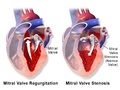 Mitral Valve Regurgitation vs Stenosis
 -
 MI Schema
 -
 Mitralinsuff TEE
 -
 Mitral Regurgitation Echo 4 Chamber
 -
 Mitral Regurgitation Echo 4 Chamber Description
 -
 Doppler Mitral Valve
 -
 The PISA Method for Quantification of Mitral Regurgitation


