Hydropneumothorax: Difference between revisions
CSV import Tags: mobile edit mobile web edit |
CSV import |
||
| Line 33: | Line 33: | ||
{{medicine-stub}} | {{medicine-stub}} | ||
{{No image}} | {{No image}} | ||
<gallery> | |||
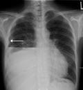
File:HydropneumoX.png|Hydropneumothorax | |||
</gallery> | |||
Revision as of 01:14, 20 February 2025
Hydropneumothorax is a medical condition characterized by the presence of both air (pneumothorax) and fluid (hydrothorax) in the pleural cavity, the space between the lung and the chest wall. This condition is often a complication of trauma, surgery, or lung disease.
Causes
Hydropneumothorax can be caused by a variety of factors, including:
- Trauma: This can include blunt or penetrating chest injuries, which can cause damage to the lung and allow air and fluid to enter the pleural cavity.
- Surgery: Certain surgical procedures, particularly those involving the lungs or chest, can inadvertently lead to hydropneumothorax.
- Lung disease: Conditions such as pneumonia, tuberculosis, and lung cancer can cause hydropneumothorax.
Symptoms
The symptoms of hydropneumothorax can vary depending on the amount of air and fluid in the pleural cavity. Common symptoms include:
Diagnosis
Hydropneumothorax is typically diagnosed through a combination of physical examination and imaging studies. A chest X-ray or CT scan can reveal the presence of air and fluid in the pleural cavity.
Treatment
The primary treatment for hydropneumothorax is to remove the air and fluid from the pleural cavity. This is typically done through a procedure called a thoracentesis, in which a needle is inserted into the pleural cavity to drain the air and fluid. In severe cases, a chest tube may be inserted to allow for continuous drainage.
See also

-
 Hydropneumothorax

