Barton's fracture: Difference between revisions
CSV import |
CSV import |
||
| Line 35: | Line 35: | ||
[[Category:Injuries]] | [[Category:Injuries]] | ||
{{Medicine-stub}} | {{Medicine-stub}} | ||
<gallery> | |||
File:3D-rendered CT of Barton's fracture.jpg|3D-rendered CT of Barton's fracture | |||
File:Radiograph of Barton's fracture.jpg|Radiograph of Barton's fracture | |||
</gallery> | |||
Revision as of 00:35, 20 February 2025
Barton's fracture is a fracture of the distal radius bone in the forearm, specifically characterized by a fracture at the base of the distal radius with dislocation of the radiocarpal joint. This injury is named after the British surgeon, John Rhea Barton, who first described it in 1838. Barton's fractures are classified into two main types based on the direction of the dislocation: dorsal Barton's fracture, where the dislocation is towards the back of the hand, and volar Barton's fracture, with dislocation towards the palm.
Causes
Barton's fractures typically occur from a fall on an outstretched hand (FOOSH injury), with the wrist in extension or flexion, depending on the type of Barton's fracture. The force of the impact causes a shearing effect at the distal radius, leading to the fracture and subsequent dislocation.
Symptoms
Common symptoms of Barton's fracture include:
- Severe pain immediately after the injury
- Swelling and bruising around the wrist
- Deformity of the wrist, indicating dislocation
- Limited range of motion in the affected wrist
Diagnosis
Diagnosis of Barton's fracture involves a thorough medical history and physical examination, followed by imaging studies. X-rays of the wrist are the primary diagnostic tool, often showing the fracture and any associated dislocation. In some cases, a CT scan may be necessary to assess the extent of the injury and plan for surgery.
Treatment
Treatment of Barton's fracture depends on the severity of the fracture and dislocation. Options include:
- Non-surgical treatment: For less severe fractures, treatment may involve immobilization of the wrist in a cast or splint for several weeks, followed by physical therapy to restore function.
- Surgical treatment: Severe fractures or those with significant dislocation may require surgery to realign the bones and fix them in place with pins, screws, or plates. Surgery is often followed by a period of immobilization and rehabilitation.
Rehabilitation
Rehabilitation is a crucial part of recovery from Barton's fracture, aiming to restore the range of motion, strength, and function of the wrist. Physical therapy exercises begin gradually and increase in intensity as the healing process progresses.
Complications
Possible complications of Barton's fracture include:
- Chronic pain
- Stiffness and reduced range of motion in the wrist
- Post-traumatic arthritis
- Nerve damage
Prevention
Preventing Barton's fractures involves minimizing the risk of falls and injuries to the wrist. This can include using protective gear during sports, improving balance and coordination through exercise, and ensuring safe environments to reduce the risk of falls.

-
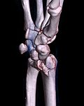 3D-rendered CT of Barton's fracture
 -
 Radiograph of Barton's fracture


