Pulmonary contusion: Difference between revisions
CSV import |
CSV import |
||
| Line 35: | Line 35: | ||
{{medicine-stub}} | {{medicine-stub}} | ||
<gallery> | |||
File:Pulmonary_contusion_CT_arrow.jpg|Pulmonary contusion CT scan with arrow | |||
File:Alveolus_diagram.svg|Diagram of an alveolus | |||
File:Fluid-filled_alveolus.svg|Fluid-filled alveolus | |||
File:Pulmonary_contusion.jpg|Pulmonary contusion | |||
File:Pulmonary_contusion_pseudocyst_CT.jpg|Pulmonary contusion pseudocyst CT | |||
File:Lung_Contusion.png|Lung contusion | |||
File:Healed_pulmonary_contusion.JPG|Healed pulmonary contusion | |||
File:AARDS_X-ray_cropped.jpg|Acute respiratory distress syndrome X-ray | |||
File:Pneumothorax_hemothorax_pneumomediastinum_contusion.JPG|Pneumothorax, hemothorax, pneumomediastinum, and contusion | |||
</gallery> | |||
Revision as of 12:22, 18 February 2025
Pulmonary contusion is a medical condition characterized by the bruising of lung tissue resulting from blunt chest trauma. This injury leads to bleeding and fluid accumulation in the lung tissue, which can significantly impair gas exchange and potentially lead to respiratory failure. Pulmonary contusions are most commonly associated with accidents such as vehicle collisions, falls from significant heights, or direct blows to the chest. Early diagnosis and management are crucial to prevent complications such as pneumonia and acute respiratory distress syndrome (ARDS).
Causes
Pulmonary contusions are primarily caused by blunt trauma to the chest. This can occur during vehicle collisions, where the chest is rapidly compressed between the seat belt and the spine, during sports injuries, or as a result of a fall. The force of the impact causes damage to the lung tissues, leading to hemorrhage and edema.
Symptoms
The symptoms of a pulmonary contusion can vary depending on the severity of the injury but typically include:
- Chest pain
- Cough, which may produce bloody or frothy sputum
- Dyspnea (difficulty breathing)
- Rapid breathing (tachypnea)
- Cyanosis (bluish discoloration of the skin due to lack of oxygen)
Diagnosis
Diagnosis of a pulmonary contusion is primarily based on the patient's history of trauma and clinical symptoms. Imaging studies play a crucial role in confirming the diagnosis and assessing the extent of the injury. The most commonly used imaging modalities include:
- Chest X-ray: May show irregular opacities in the lung fields, although findings might not be apparent immediately after the injury.
- Computed tomography (CT) scan: More sensitive than a chest X-ray and can detect smaller contusions, showing detailed images of the lung tissue.
Treatment
The treatment of pulmonary contusion focuses on supporting the patient's breathing and preventing complications. Treatment strategies may include:
- Oxygen therapy to maintain adequate oxygen levels in the blood.
- Pain management to enable deep breathing and coughing, which are essential to clear the lungs.
- In severe cases, mechanical ventilation may be required to support the patient's breathing.
- Monitoring and treatment for complications such as pneumonia or ARDS.
Prevention
Preventing accidents that lead to chest trauma is the best way to prevent pulmonary contusions. This includes wearing seat belts, using appropriate protective gear during sports, and ensuring safety measures are in place when working at heights.
Prognosis
The prognosis for individuals with pulmonary contusion depends on the severity of the injury and the presence of associated injuries. Most patients recover fully with appropriate treatment, but severe cases can be life-threatening.

-
 Pulmonary contusion CT scan with arrow
 -
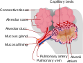 Diagram of an alveolus
 -
 Fluid-filled alveolus
 -
 Pulmonary contusion
 -
 Pulmonary contusion pseudocyst CT
 -
 Lung contusion
 -
 Healed pulmonary contusion
 -
 Acute respiratory distress syndrome X-ray
 -
Pneumothorax, hemothorax, pneumomediastinum, and contusion


