Artistic gymnastics: Difference between revisions
CSV import |
CSV import |
||
| Line 31: | Line 31: | ||
[[Category:Neuroanatomy]] | [[Category:Neuroanatomy]] | ||
<gallery> | |||
File:Illia_Kovtun_performing_in_the_men's_parallel_bars_final_at_Paris_2024.png|Artistic gymnastics | |||
File:Bundesarchiv_Bild_183-94681-0002,_Werner_Dölling.jpg|Artistic gymnastics | |||
File:DHypolito-Vault.jpg|Artistic gymnastics | |||
File:2018-10-14_Gymnastics_at_2018_Summer_Youth_Olympics_–_Boys'_Artistic_Gymnastics_–_Apparatus_finals_–_Vault_(Martin_Rulsch)_176.jpg|Artistic gymnastics | |||
File:2015_European_Artistic_Gymnastics_Championships_-_Vault_-_Ksenia_Afanasyeva_08.jpg|Artistic gymnastics | |||
File:EUA_levam_ouro_na_ginástica_artística_feminina;_Brasil_fica_em_8º_lugar_(28264942923).jpg|Artistic gymnastics | |||
File:2019-06-29_1st_FIG_Artistic_Gymnastics_JWCH_Men's_Apparatus_finals_Floor_exercise_(Martin_Rulsch)_420.jpg|Artistic gymnastics | |||
File:Polina_Astakhova_1960.jpg|Artistic gymnastics | |||
File:Alberto_Braglia.jpg|Artistic gymnastics | |||
File:2015_European_Artistic_Gymnastics_Championships_-_Pommel_horse_-_Louis_Smith_06.jpg|Artistic gymnastics | |||
File:De_Russische_Michael_Woronine_in_aktie_op_het_paard,_Bestanddeelnr_919-5874.jpg|Artistic gymnastics | |||
File:Bundesarchiv_Bild_183-N0621-0038,_DDR-Meisterschaften_im_Turnen,_Klaus_Köste.jpg|Artistic gymnastics | |||
</gallery> | |||
Latest revision as of 12:07, 18 February 2025
Arachnoid Trabeculae[edit]
The arachnoid trabeculae are delicate, web-like structures that are part of the arachnoid mater, one of the three layers of the meninges that cover the central nervous system. These trabeculae extend through the subarachnoid space, connecting the arachnoid mater to the underlying pia mater.
Structure[edit]
The arachnoid trabeculae are composed of collagen fibers and fibroblasts, which provide structural support and maintain the spacing between the arachnoid and pia mater. This spacing is crucial for the circulation of cerebrospinal fluid (CSF) within the subarachnoid space. The trabeculae are most dense in areas where the brain is subject to movement, providing additional support and stability.
Function[edit]
The primary function of the arachnoid trabeculae is to maintain the subarachnoid space, allowing for the free flow of CSF. This fluid acts as a cushion, protecting the brain and spinal cord from mechanical injury. Additionally, the trabeculae help to anchor the brain within the cranial cavity, preventing excessive movement that could lead to injury.
Clinical Significance[edit]
Disruption or damage to the arachnoid trabeculae can lead to various medical conditions. For example, subarachnoid hemorrhage can occur if blood vessels within the subarachnoid space are ruptured, potentially affecting the trabeculae. In some cases, arachnoid trabeculae may become thickened or scarred, leading to conditions such as arachnoiditis, which can cause chronic pain and neurological deficits.
Research[edit]
Recent studies have focused on the role of arachnoid trabeculae in the pathophysiology of hydrocephalus, a condition characterized by an abnormal accumulation of CSF. Understanding the mechanical properties and biological functions of the trabeculae may provide insights into new therapeutic approaches for managing this condition.
Related Pages[edit]
Gallery[edit]
-
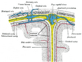 Diagram of arachnoid trabeculae within the subarachnoid space.

-
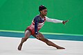
 Artistic gymnastics
 -
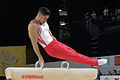
 Artistic gymnastics
 -
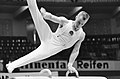
 Artistic gymnastics
 -
 Artistic gymnastics
 -
 Artistic gymnastics
 -
 Artistic gymnastics
 -
 Artistic gymnastics
 -
 Artistic gymnastics
 -
 Artistic gymnastics
 -
 Artistic gymnastics
 -
 Artistic gymnastics
 -
 Artistic gymnastics


_176.jpg)

.jpg)
_420.jpg)


